Movie
Movie Controller

[English] 日本語
 Yorodumi
- EMDB-47728: Structure of thioferritin (PfDPSL) with ferrihydrite growth at a ... -
+ Open data

- Basic information
| Entry |  | |||||||||
|---|---|---|---|---|---|---|---|---|---|---|
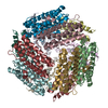
| Title | Structure of thioferritin (PfDPSL) with ferrihydrite growth at a single three-fold pore. | |||||||||
 Map data Map data | Sharpened map | |||||||||
 Sample Sample |
| |||||||||
 Keywords Keywords | Ferritin / thioferritin / oxidative stress / iron homeostasis / iron mineral / ferric oxyhydroxide / mineral core / protein cage / METAL BINDING PROTEIN | |||||||||
| Function / homology |  Function and homology information Function and homology informationOxidoreductases; Oxidizing metal ions / nucleoid / ferroxidase activity / ferric iron binding / heme binding / DNA binding / cytosol Similarity search - Function | |||||||||
| Biological species |   Pyrococcus furiosus (archaea) Pyrococcus furiosus (archaea) | |||||||||
| Method | single particle reconstruction / cryo EM / Resolution: 2.48 Å | |||||||||
 Authors Authors | Gauvin CC / Waghwani HK / Tokmina-Lukaszewska M / Bothner B / Douglas T / Lawrence CM | |||||||||
| Funding support |  United States, 1 items United States, 1 items
| |||||||||
 Citation Citation | Journal: J Am Chem Soc / Year: 2025 Title: The Mechanism of Mineral Nucleation and Growth in a Mini-Ferritin. Authors: Colin C Gauvin / Monika Tokmina-Lukaszewska / Hitesh Kumar Waghwani / Sterling C McBee / Trevor Douglas / Brian Bothner / C Martin Lawrence / Abstract: Iron is an enigmatic element. While necessary for life, as Fe(II) it also catalyzes formation of reactive oxygen species. To mitigate this, cellular life has evolved the ferritin protein superfamily, ...Iron is an enigmatic element. While necessary for life, as Fe(II) it also catalyzes formation of reactive oxygen species. To mitigate this, cellular life has evolved the ferritin protein superfamily, which includes the 24 subunit ferritins and bacterioferritins, and 12 subunit mini-ferritins (DPS). Each catalyze the oxidation of Fe(II) to ferric oxyhydroxide, which is then sequestered within the hollow protein shell. While there is a wealth of structural information on unmineralized ferritins, high resolution information on iron loaded ferritins is lacking, and the mechanism of iron mineralization is poorly understood. To address this, we followed iron loading in a mini-ferritin by cryo-EM. We determined a 1.86 Å structure in the unmineralized state, as well as a 1.91 Å structure of an early, iron loading state in which the mini-ferritin catalyzes nucleation of ferric oxyhydroxide at the acidic 3-fold pores. Mechanistically, a conserved crucible of precisely positioned glutamates and unsaturated main chain carbonyls are employed as a template to catalyze nucleation. A 2.4 Å structure at a later time point was also determined, revealing the role of a second constellation of main-chain carbonyls on the interior surface that subsequently supports crystalline mineral growth, that then proceeds into the center of the particle. Notably, the visualized mineral is consistent with one of two competing structural descriptions for ferrihydrite. This study provides the first pseudoatomic level observation of controlled mineral nucleation and growth in any member of the ferritin superfamily, and informs general mechanisms of nucleation and biomineralization. | |||||||||
| History |
|
- Structure visualization
| Supplemental images |
|---|

- Downloads & links
-EMDB archive
| Map data | emd_47728.map.gz | 322.8 MB | EMDB map data format | |
|---|---|---|---|---|
| Header (meta data) | emd-47728-v30.xmlemd-47728.xml | 23.7 KB 23.7 KB | Display Display | EMDB header |
| FSC (resolution estimation) | emd_47728_fsc.xml | 14.8 KB | Display | FSC data file |
| Images |  emd_47728.png emd_47728.png | 54.8 KB | ||
| Filedesc metadata | emd-47728.cif.gz | 7 KB | ||
| Others | emd_47728_half_map_1.map.gzemd_47728_half_map_2.map.gz | 318 MB 318 MB | ||
| Archive directory |  http://ftp.pdbj.org/pub/emdb/structures/EMD-47728ftp://ftp.pdbj.org/pub/emdb/structures/EMD-47728 http://ftp.pdbj.org/pub/emdb/structures/EMD-47728ftp://ftp.pdbj.org/pub/emdb/structures/EMD-47728 | HTTPS FTP |
-Related structure data
| Related structure data |  9e8sMC  9cz0C  9cz8C  9cz9C M: atomic model generated by this map C: citing same article ( |
|---|---|
| Similar structure data |


-Links
| EMDB pages | EMDB (EBI/PDBe) / EMDataResource |
|---|---|
| Related items in Molecule of the Month |


-Map
| File | Download / File: emd_47728.map.gz / Format: CCP4 / Size: 343 MB / Type: IMAGE STORED AS FLOATING POINT NUMBER (4 BYTES) | ||||||||||||||||||||||||||||||||||||
|---|---|---|---|---|---|---|---|---|---|---|---|---|---|---|---|---|---|---|---|---|---|---|---|---|---|---|---|---|---|---|---|---|---|---|---|---|---|
| Annotation | Sharpened map | ||||||||||||||||||||||||||||||||||||
| Projections & slices | Image control
Images are generated by Spider. | ||||||||||||||||||||||||||||||||||||
| Voxel size | X=Y=Z: 0.69 Å | ||||||||||||||||||||||||||||||||||||
| Density |
| ||||||||||||||||||||||||||||||||||||
| Symmetry | Space group: 1 | ||||||||||||||||||||||||||||||||||||
| Details | EMDB XML:
|
 Z (Sec.)
 Y (Row.)
 X (Col.)


-Supplemental data
-Half map: Half map B
| File | emd_47728_half_map_1.map | ||||||||||||
|---|---|---|---|---|---|---|---|---|---|---|---|---|---|
| Annotation | Half map B | ||||||||||||
| Projections & Slices |
| ||||||||||||
| Density Histograms |


-Half map: Half map A
| File | emd_47728_half_map_2.map | ||||||||||||
|---|---|---|---|---|---|---|---|---|---|---|---|---|---|
| Annotation | Half map A | ||||||||||||
| Projections & Slices |
| ||||||||||||
| Density Histograms |


- Sample components
-Entire : Thioferritin dodecamer with loaded ferroxidase centers and minera...
| Entire | Name: Thioferritin dodecamer with loaded ferroxidase centers and mineral at single 3-fold pore |
|---|---|
| Components |
|
-Supramolecule #1: Thioferritin dodecamer with loaded ferroxidase centers and minera...
| Supramolecule | Name: Thioferritin dodecamer with loaded ferroxidase centers and mineral at single 3-fold pore type: complex / ID: 1 / Parent: 0 / Macromolecule list: #1 |
|---|---|
| Source (natural) | Organism: Pyrococcus furiosus (archaea) |
| Molecular weight | Theoretical: 256 KDa |
-Macromolecule #1: DNA protection during starvation protein
| Macromolecule | Name: DNA protection during starvation protein / type: protein_or_peptide / ID: 1 / Number of copies: 12 / Enantiomer: LEVO / EC number: Oxidoreductases; Oxidizing metal ions |
|---|---|
| Source (natural) | Organism: Pyrococcus furiosus (archaea) |
| Molecular weight | Theoretical: 21.387254 KDa |
| Recombinant expression | Organism:  |
| Sequence | String: MPEHNRRLVE RTGIDVEKLL ELLIKAAAAE FTTYYYYTIL RNHATGLEGE AIKEIIEDAR LEDRNHFEAL VPRIYELGGE LPRDIREFA DLASCRDAYL PEEPTIENIL KVLLEAERCA VGVYTEICNY TFGKDPRTYD LALAILHEEI EHEAWFEELL T GKPSGHFR RGKPGESPYV SKFLKTR UniProtKB: DNA protection during starvation protein |
-Macromolecule #2: FE (III) ION
| Macromolecule | Name: FE (III) ION / type: ligand / ID: 2 / Number of copies: 37 / Formula: FE |
|---|---|
| Molecular weight | Theoretical: 55.845 Da |
-Macromolecule #3: OXYGEN MOLECULE
| Macromolecule | Name: OXYGEN MOLECULE / type: ligand / ID: 3 / Number of copies: 12 / Formula: OXY |
|---|---|
| Molecular weight | Theoretical: 31.999 Da |
| Chemical component information |  ChemComp-O2: |
-Macromolecule #4: OXYGEN ATOM
| Macromolecule | Name: OXYGEN ATOM / type: ligand / ID: 4 / Number of copies: 18 / Formula: O |
|---|---|
| Molecular weight | Theoretical: 15.999 Da |
| Chemical component information |  ChemComp-O: |
-Macromolecule #5: water
| Macromolecule | Name: water / type: ligand / ID: 5 / Number of copies: 144 / Formula: HOH |
|---|---|
| Molecular weight | Theoretical: 18.015 Da |
| Chemical component information |  ChemComp-HOH: |
-Experimental details
-Structure determination
| Method | cryo EM |
|---|---|
 Processing Processing | single particle reconstruction |
| Aggregation state | particle |
-Sample preparation
| Concentration | 2.5 mg/mL | |||||||||
|---|---|---|---|---|---|---|---|---|---|---|
| Buffer | pH: 6.5 Component:
Details: 50 mM MES, 100 mM NaCl, pH 6.5 | |||||||||
| Grid | Model: Quantifoil / Material: COPPER / Mesh: 300 / Support film - Material: CARBON / Support film - topology: HOLEY / Pretreatment - Type: GLOW DISCHARGE / Pretreatment - Time: 45 sec. / Pretreatment - Atmosphere: AIR | |||||||||
| Vitrification | Cryogen name: ETHANE / Chamber humidity: 95 % / Chamber temperature: 277.15 K / Instrument: FEI VITROBOT MARK IV | |||||||||
| Details | This sample formed a thin monodispersed layer. |
- Electron microscopy
| Microscope | FEI TALOS ARCTICA |
|---|---|
| Image recording | Film or detector model: GATAN K3 (6k x 4k) / Number grids imaged: 1 / Number real images: 9094 / Average exposure time: 6.0 sec. / Average electron dose: 55.0 e/Å2 |
| Electron beam | Acceleration voltage: 200 kV / Electron source:  FIELD EMISSION GUN FIELD EMISSION GUN |
| Electron optics | C2 aperture diameter: 50.0 µm / Illumination mode: FLOOD BEAM / Imaging mode: BRIGHT FIELD / Cs: 2.7 mm / Nominal defocus max: 1.6 µm / Nominal defocus min: 0.6 µm / Nominal magnification: 57000 |
| Sample stage | Specimen holder model: FEI TITAN KRIOS AUTOGRID HOLDER / Cooling holder cryogen: NITROGEN |
| Experimental equipment |  Model: Talos Arctica / Image courtesy: FEI Company |
+Image processing

-Atomic model buiding 1
| Initial model | PDB ID: Chain - Source name: PDB / Chain - Initial model type: experimental model |
|---|---|
| Details | Model was fit to map using ChimeraX fitmap, and then refined in PHENIX. |
| Refinement | Space: REAL / Protocol: RIGID BODY FIT / Overall B value: 58.4 / Target criteria: Cross-correlation coefficient |
| Output model | PDB-9e8s: |