National Institutes of Health/National Cancer Institute (NIH/NCI)
United States
Citation
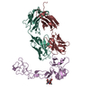
Journal: Biomed Pharmacother / Year: 2024 Title: Fully human monoclonal antibody targeting the cysteine-rich substrate-interacting region of ADAM17 on cancer cells. Authors: Nayanendu Saha / Sang Gyu Lee / Eeva-Christine Brockmann / M Jason de la Cruz / Yehuda Goldgur / Rachelle P Mendoza / Elisa de Stanchina / Tanzy M Love / Josh Marvald / Yan Xu / Kai Xu / ...Authors: Nayanendu Saha / Sang Gyu Lee / Eeva-Christine Brockmann / M Jason de la Cruz / Yehuda Goldgur / Rachelle P Mendoza / Elisa de Stanchina / Tanzy M Love / Josh Marvald / Yan Xu / Kai Xu / Juha P Himanen / Urpo Lamminmäki / Darren Veach / Dimitar B Nikolov / Abstract: ADAM17 sheds EGFR/erbB ligands and triggers oncogenic pathways that lead to the progression of solid tumors. We targeted the ADAM17 disintegrin and cysteine rich domain region (D+C) to generate a ...ADAM17 sheds EGFR/erbB ligands and triggers oncogenic pathways that lead to the progression of solid tumors. We targeted the ADAM17 disintegrin and cysteine rich domain region (D+C) to generate a panel of single-chain antibody fragments (scFvs) that selectively bind to the D or C domains of ADAM17, but not of ADAM10 or ADAM19. From the panel, we selected one scFv, referred to as C12, based on its high binding affinity towards the target, and re-formatted it to a full IgG for further studies. High-resolution cryo-electron microscopy studies documented that the mAb binds to the ADAM17 C-domain that in ADAM proteases, notably ADAM10 and ADAM17, is known to impart substrate-specificity. The C12 mAb significantly inhibited EGFR phosphorylation in cancer cell lines by hindering the cleavage of EGFR ligands tethered to the cell surface. This inhibition provides a mechanism for potential anti-tumor effects, and indeed C12 diminished the viability of a variety of EGFR-expressing cancer cell lines. Cell-based ELISA studies revealed that C12 preferentially bound to activated ADAM17 present on tumor cells, as compared to the autoinhibited ADAM17 that is the predominant form on HEK293 and other non-tumor cells. C12 also exhibited tumor growth inhibition in an ovarian cancer xenograft mouse model. Consistent with its selective tumor cell binding in vitro, radioimmuno PET (positron emission tomography) imaging with Zr-DFO-C12 in mouse xenograft models confirmed tumoral accumulation of the C12 mAb.
In the structure databanks used in Yorodumi, some data are registered as the other names, "COVID-19 virus" and "2019-nCoV". Here are the details of the virus and the list of structure data.
Jan 31, 2019. EMDB accession codes are about to change! (news from PDBe EMDB page)
EMDB accession codes are about to change! (news from PDBe EMDB page)
The allocation of 4 digits for EMDB accession codes will soon come to an end. Whilst these codes will remain in use, new EMDB accession codes will include an additional digit and will expand incrementally as the available range of codes is exhausted. The current 4-digit format prefixed with “EMD-” (i.e. EMD-XXXX) will advance to a 5-digit format (i.e. EMD-XXXXX), and so on. It is currently estimated that the 4-digit codes will be depleted around Spring 2019, at which point the 5-digit format will come into force.
The EM Navigator/Yorodumi systems omit the EMD- prefix.
Related info.:Q: What is EMD? / ID/Accession-code notation in Yorodumi/EM Navigator
Yorodumi is a browser for structure data from EMDB, PDB, SASBDB, etc.
This page is also the successor to EM Navigator detail page, and also detail information page/front-end page for Omokage search.
The word "yorodu" (or yorozu) is an old Japanese word meaning "ten thousand". "mi" (miru) is to see.
Related info.:EMDB / PDB / SASBDB / Comparison of 3 databanks / Yorodumi Search / Aug 31, 2016. New EM Navigator & Yorodumi / Yorodumi Papers / Jmol/JSmol / Function and homology information / Changes in new EM Navigator and Yorodumi
 Movie
Movie Controller

 Yorodumi
 Open data
 Basic information
 Map data
 Sample
 Keywords
 Function and homology information
 Homo sapiens (human)
 Authors
 United States, 1 items
  Citation
 Structure visualization
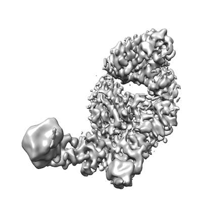
 Downloads & links
 emd_47571.png
 http://ftp.pdbj.org/pub/emdb/structures/EMD-47571
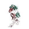


 X (Sec.)
 Y (Row.)
 Z (Col.)


 Sample components
 Spodoptera (butterflies/moths)
 Processing
 Electron microscopy
 FIELD EMISSION GUN