 Movie
Movie Controller


+ Open data

- Basic information
| Entry |  | |||||||||
|---|---|---|---|---|---|---|---|---|---|---|
| Title | Escherichia coli encapsulin-associated DyP peroxidase | |||||||||
 Map data Map data | ||||||||||
 Sample Sample |
| |||||||||
 Keywords Keywords | Dye-decolorizing peroxidase / peroxidase / DyP / encapsulin / OXIDOREDUCTASE | |||||||||
| Function / homology |  Function and homology information Function and homology information | |||||||||
| Biological species |  | |||||||||
| Method | single particle reconstruction / cryo EM / Resolution: 3.31 Å | |||||||||
 Authors Authors | Andreas MP / Ubilla NC / Giessen TW | |||||||||
| Funding support |  United States, 1 items United States, 1 items
| |||||||||
 Citation Citation | Journal: bioRxiv / Year: 2024 Title: Structural and biochemical characterization of a widespread enterobacterial peroxidase encapsulin. Authors: Natalia C Ubilla-Rodriguez / Michael P Andreas / Tobias W Giessen / Abstract: Encapsulins are self-assembling protein compartments found in prokaryotes and specifically encapsulate dedicated cargo enzymes. The most abundant encapsulin cargo class are Dye-decolorizing ...Encapsulins are self-assembling protein compartments found in prokaryotes and specifically encapsulate dedicated cargo enzymes. The most abundant encapsulin cargo class are Dye-decolorizing Peroxidases (DyPs). It has been previously suggested that DyP encapsulins are involved in oxidative stress resistance and bacterial pathogenicity due to DyPs' inherent ability to reduce and detoxify hydrogen peroxide while oxidizing a broad range of organic co-substrates. Here, we report the structural and biochemical analysis of a DyP encapsulin widely found across enterobacteria. Using bioinformatic approaches, we show that this DyP encapsulin is encoded by a conserved transposon-associated operon, enriched in enterobacterial pathogens. Through low pH and peroxide exposure experiments, we highlight the stability of this DyP encapsulin under harsh conditions and show that DyP catalytic activity is highest at low pH. We determine the structure of the DyP-loaded shell and free DyP via cryo-electron microscopy, revealing the structural basis for DyP cargo loading and peroxide preference. Our work lays the foundation to further explore the substrate range and physiological functions of enterobacterial DyP encapsulins. | |||||||||
| History |
|
- Structure visualization
| Supplemental images |
|---|

- Downloads & links
-EMDB archive
| Map data | emd_47518.map.gz | 59.3 MB | EMDB map data format | |
|---|---|---|---|---|
| Header (meta data) | emd-47518-v30.xmlemd-47518.xml | 20.3 KB 20.3 KB | Display Display | EMDB header |
| FSC (resolution estimation) | emd_47518_fsc.xml | 8.5 KB | Display | FSC data file |
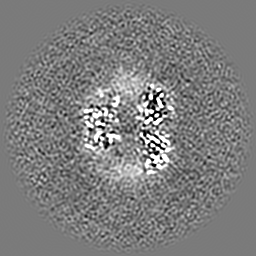
| Images |  emd_47518.png emd_47518.png | 107.7 KB | ||
| Filedesc metadata | emd-47518.cif.gz | 6.6 KB | ||
| Others | emd_47518_half_map_1.map.gzemd_47518_half_map_2.map.gz | 59.2 MB 59.2 MB | ||
| Archive directory |  http://ftp.pdbj.org/pub/emdb/structures/EMD-47518ftp://ftp.pdbj.org/pub/emdb/structures/EMD-47518 http://ftp.pdbj.org/pub/emdb/structures/EMD-47518ftp://ftp.pdbj.org/pub/emdb/structures/EMD-47518 | HTTPS FTP |
-Related structure data
| Related structure data |  9e4rMC  9e5eC M: atomic model generated by this map C: citing same article ( |
|---|---|
| Similar structure data |

-Links
| EMDB pages | EMDB (EBI/PDBe) / EMDataResource |
|---|
-Map
| File | Download / File: emd_47518.map.gz / Format: CCP4 / Size: 64 MB / Type: IMAGE STORED AS FLOATING POINT NUMBER (4 BYTES) | ||||||||||||||||||||||||||||||||||||
|---|---|---|---|---|---|---|---|---|---|---|---|---|---|---|---|---|---|---|---|---|---|---|---|---|---|---|---|---|---|---|---|---|---|---|---|---|---|
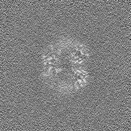
| Projections & slices | Image control
Images are generated by Spider. | ||||||||||||||||||||||||||||||||||||
| Voxel size | X=Y=Z: 0.91 Å | ||||||||||||||||||||||||||||||||||||
| Density |
| ||||||||||||||||||||||||||||||||||||
| Symmetry | Space group: 1 | ||||||||||||||||||||||||||||||||||||
| Details | EMDB XML:
|
 Z (Sec.)
 X (Row.)
 Y (Col.)


-Supplemental data
-Half map: #2
| File | emd_47518_half_map_1.map | ||||||||||||
|---|---|---|---|---|---|---|---|---|---|---|---|---|---|
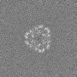
| Projections & Slices |
| ||||||||||||
| Density Histograms |


-Half map: #1
| File | emd_47518_half_map_2.map | ||||||||||||
|---|---|---|---|---|---|---|---|---|---|---|---|---|---|
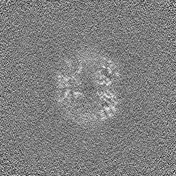
| Projections & Slices |
| ||||||||||||
| Density Histograms |


- Sample components
-Entire : Escherichia coli KTE40 encapsulin-associated DyP peroxidase
| Entire | Name: Escherichia coli KTE40 encapsulin-associated DyP peroxidase |
|---|---|
| Components |
|
-Supramolecule #1: Escherichia coli KTE40 encapsulin-associated DyP peroxidase
| Supramolecule | Name: Escherichia coli KTE40 encapsulin-associated DyP peroxidase type: complex / ID: 1 / Parent: 0 / Macromolecule list: #1 |
|---|---|
| Source (natural) | Organism: |
-Macromolecule #1: Dyp-type peroxidase
| Macromolecule | Name: Dyp-type peroxidase / type: protein_or_peptide / ID: 1 / Number of copies: 1 / Enantiomer: LEVO |
|---|---|
| Source (natural) | Organism: |
| Molecular weight | Theoretical: 40.644516 KDa |
| Recombinant expression | Organism: |
| Sequence | String: MHHHHHHGGS ENLYFQSGGS GCPFSQSVSQ PVDERLTRAA IFLVVTINPG KAAEVAVRAH CSILSSLIRG VGFRISDGGL SCVMGVSEG GWERLFGDTK PEYLHVFREI NGVHHAPSTP GDLLYHIRAA RMDLCFELAS RILSDLGNSV SVVDSVQGFR Y FDDRDLLG ...String: MHHHHHHGGS ENLYFQSGGS GCPFSQSVSQ PVDERLTRAA IFLVVTINPG KAAEVAVRAH CSILSSLIRG VGFRISDGGL SCVMGVSEG GWERLFGDTK PEYLHVFREI NGVHHAPSTP GDLLYHIRAA RMDLCFELAS RILSDLGNSV SVVDSVQGFR Y FDDRDLLG FVDGTENPVA QAAVDATLIG DEDMVFAGGS YVIVQKYLHD LDKWNAIPVE QQEKIIGREK LSDIELRDAD KP SYAHNVL TSIEEDGEDV DILRDNMPFG DPGKGEFGTY FIGYSRKPER IERMLENMFI GNPPGNYDRI LDVSRAITGT LFF VPTTSF LDSIEPQSAP GQQGDDVINT LRSTAIKGDI MPGSLNIGSL KKEV UniProtKB: Dyp-type peroxidase |
-Macromolecule #2: PROTOPORPHYRIN IX CONTAINING FE
| Macromolecule | Name: PROTOPORPHYRIN IX CONTAINING FE / type: ligand / ID: 2 / Number of copies: 1 / Formula: HEM |
|---|---|
| Molecular weight | Theoretical: 616.487 Da |
| Chemical component information |  ChemComp-HEM: |
-Experimental details
-Structure determination
| Method | cryo EM |
|---|---|
 Processing Processing | single particle reconstruction |
| Aggregation state | particle |
-Sample preparation
| Concentration | 0.5 mg/mL | |||||||||
|---|---|---|---|---|---|---|---|---|---|---|
| Buffer | pH: 7.5 Component:
Details: Sample was prepared in 20 mM Tris pH 7.5, 150 mM NaCl. | |||||||||
| Grid | Model: Quantifoil R1.2/1.3 / Material: COPPER / Mesh: 200 / Support film - Material: CARBON / Support film - topology: HOLEY / Pretreatment - Type: GLOW DISCHARGE / Pretreatment - Time: 60 sec. Details: Grids were glow discharged for 60 seconds at 5 mA in vacuum. | |||||||||
| Vitrification | Cryogen name: ETHANE / Chamber humidity: 100 % / Chamber temperature: 295 K / Instrument: FEI VITROBOT MARK IV Details: Grids were frozen with the following parameters: blot force 20, blot time 4 seconds, humidty 100 percent, 22 degrees C.. | |||||||||
| Details | Sample was frozen on two grids, one at 0.5 mg/mL and the other at 0.25 mg/mL. |
- Electron microscopy #1
| Microscopy ID | 1 |
|---|---|
| Microscope | FEI TALOS ARCTICA |
| Image recording | Image recording ID: 1 / Film or detector model: GATAN K2 SUMMIT (4k x 4k) / Detector mode: COUNTING / Digitization - Dimensions - Width: 3710 pixel / Digitization - Dimensions - Height: 3838 pixel / Number grids imaged: 1 / Number real images: 123 / Average exposure time: 6.0 sec. / Average electron dose: 47.84 e/Å2 |
| Electron beam | Acceleration voltage: 200 kV / Electron source:  FIELD EMISSION GUN FIELD EMISSION GUN |
| Electron optics | Illumination mode: FLOOD BEAM / Imaging mode: BRIGHT FIELD / Cs: 2.7 mm / Nominal defocus max: 1.8 µm / Nominal defocus min: 1.0 µm / Nominal magnification: 45000 |
| Sample stage | Cooling holder cryogen: NITROGEN |
| Experimental equipment |  Model: Talos Arctica / Image courtesy: FEI Company |
-Electron microscopy #1~
| Microscopy ID | 1 |
|---|---|
| Microscope | TFS GLACIOS |
| Image recording | Image recording ID: 2 / Film or detector model: GATAN K2 SUMMIT (4k x 4k) / Detector mode: COUNTING / Digitization - Dimensions - Width: 3710 pixel / Digitization - Dimensions - Height: 3838 pixel / Number grids imaged: 1 / Number real images: 206 / Average exposure time: 4.0 sec. / Average electron dose: 46.24 e/Å2 |
| Electron beam | Acceleration voltage: 200 kV / Electron source: FIELD EMISSION GUN |
| Electron optics | Illumination mode: FLOOD BEAM / Imaging mode: BRIGHT FIELD / Cs: 2.7 mm / Nominal defocus max: 1.8 µm / Nominal defocus min: 1.0 µm / Nominal magnification: 45000 |


