Movie
Movie Controller


+ Open data

- Basic information
| Entry |  | |||||||||
|---|---|---|---|---|---|---|---|---|---|---|
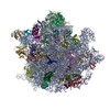
| Title | M. smegmatis methylated 70S ribosome structure | |||||||||
 Map data Map data | Unsharpened postprocessed map of the methylated M. smegmatis 70S ribosome | |||||||||
 Sample Sample |
| |||||||||
 Keywords Keywords | 70S ribosome / unmethylated ribosome / ribonucleoprotein complex / protein synthesis / RIBOSOME | |||||||||
| Function / homology |  Function and homology information Function and homology informationlarge ribosomal subunit / transferase activity / ribosomal small subunit assembly / ribosome biogenesis / ribosomal small subunit biogenesis / 5S rRNA binding / ribosomal large subunit assembly / small ribosomal subunit / small ribosomal subunit rRNA binding / large ribosomal subunit rRNA binding ...large ribosomal subunit / transferase activity / ribosomal small subunit assembly / ribosome biogenesis / ribosomal small subunit biogenesis / 5S rRNA binding / ribosomal large subunit assembly / small ribosomal subunit / small ribosomal subunit rRNA binding / large ribosomal subunit rRNA binding / cytosolic small ribosomal subunit / cytosolic large ribosomal subunit / cytoplasmic translation / tRNA binding / negative regulation of translation / rRNA binding / structural constituent of ribosome / ribosome / translation / ribonucleoprotein complex / mRNA binding / RNA binding / zinc ion binding / metal ion binding / cytoplasm / cytosol Similarity search - Function | |||||||||
| Biological species |  Mycolicibacterium smegmatis (bacteria) Mycolicibacterium smegmatis (bacteria) | |||||||||
| Method | single particle reconstruction / cryo EM / Resolution: 3.17 Å | |||||||||
 Authors Authors | Nandi S / Conn GL | |||||||||
| Funding support |  United States, 1 items United States, 1 items
| |||||||||
 Citation Citation | Journal: Nucleic Acids Res / Year: 2025 Title: Distant ribose 2'-O-methylation of 23S rRNA helix 69 pre-orders the capreomycin drug binding pocket at the ribosome subunit interface. Authors: Suparno Nandi / Debayan Dey / Pooja Srinivas / Christine M Dunham / Graeme L Conn / Abstract: Loss of ribosomal RNA (rRNA) modifications incorporated by the intrinsic methyltransferase TlyA results in reduced sensitivity to tuberactinomycin antibiotics such as capreomycin. However, how rRNA ...Loss of ribosomal RNA (rRNA) modifications incorporated by the intrinsic methyltransferase TlyA results in reduced sensitivity to tuberactinomycin antibiotics such as capreomycin. However, how rRNA methylation alters drug binding, particularly at the distant but functionally more important site in 23S rRNA helix 69 (H69), is currently unknown. We determined high-resolution cryo-electron microscopy structures of the Mycolicibacterium smegmatis 70S ribosome with or without the two ribose 2'-O-methyl modifications incorporated by TlyA. In the unmodified ribosome, the tip of H69 adopts a more compact conformation, positioning two key nucleotides (A2137 and C2138) such that interactions with capreomycin would be lost and the binding pocket partially occluded. Methylation of 23S rRNA nucleotide C2144 promotes conformational changes that result in a more favorable positioning of C2138 and adoption of a more open conformation to enable capreomycin binding. Molecular dynamics simulations and H69 RNA helical analyses additionally reveal specific propagation of these changes from the site of modification to the H69 tip, allosterically reconfiguring the capreomycin binding site. Methylation of h44 also results in structural rearrangements at the H69-h44 interface to support maintenance of these changes that favor antibiotic binding. This work thus reveals the effect and regulation of distant rRNA methylation on ribosome-targeting antibiotic binding. | |||||||||
| History |
|
- Structure visualization
| Supplemental images |
|---|

- Downloads & links
-EMDB archive
| Map data | emd_47365.map.gz | 222.1 MB | EMDB map data format | |
|---|---|---|---|---|
| Header (meta data) | emd-47365-v30.xmlemd-47365.xml | 104.3 KB 104.3 KB | Display Display | EMDB header |
| FSC (resolution estimation) | emd_47365_fsc.xml | 14.1 KB | Display | FSC data file |
| Images |  emd_47365.png emd_47365.png | 57.8 KB | ||
| Filedesc metadata | emd-47365.cif.gz | 15.2 KB | ||
| Others | emd_47365_additional_1.map.gzemd_47365_additional_10.map.gzemd_47365_additional_11.map.gzemd_47365_additional_12.map.gzemd_47365_additional_13.map.gzemd_47365_additional_2.map.gzemd_47365_additional_3.map.gzemd_47365_additional_4.map.gzemd_47365_additional_5.map.gzemd_47365_additional_6.map.gzemd_47365_additional_7.map.gzemd_47365_additional_8.map.gzemd_47365_additional_9.map.gzemd_47365_half_map_1.map.gzemd_47365_half_map_2.map.gz | 157 MB 225 MB 219.8 MB 217.1 MB 216.8 MB 157.1 MB 177.5 MB 177.1 MB 155 MB 174.4 MB 189.7 MB 219.1 MB 225.4 MB 193.6 MB 193.9 MB | ||
| Archive directory |  http://ftp.pdbj.org/pub/emdb/structures/EMD-47365ftp://ftp.pdbj.org/pub/emdb/structures/EMD-47365 http://ftp.pdbj.org/pub/emdb/structures/EMD-47365ftp://ftp.pdbj.org/pub/emdb/structures/EMD-47365 | HTTPS FTP |
-Related structure data
| Related structure data |  9e0pMC  9e0nC M: atomic model generated by this map C: citing same article ( |
|---|---|
| Similar structure data |

-Links
| EMDB pages | EMDB (EBI/PDBe) / EMDataResource |
|---|---|
| Related items in Molecule of the Month |


-Map
| File | Download / File: emd_47365.map.gz / Format: CCP4 / Size: 244.1 MB / Type: IMAGE STORED AS FLOATING POINT NUMBER (4 BYTES) | ||||||||||||||||||||||||||||||||||||
|---|---|---|---|---|---|---|---|---|---|---|---|---|---|---|---|---|---|---|---|---|---|---|---|---|---|---|---|---|---|---|---|---|---|---|---|---|---|
| Annotation | Unsharpened postprocessed map of the methylated M. smegmatis 70S ribosome | ||||||||||||||||||||||||||||||||||||
| Projections & slices | Image control
Images are generated by Spider. | ||||||||||||||||||||||||||||||||||||
| Voxel size | X=Y=Z: 1.069 Å | ||||||||||||||||||||||||||||||||||||
| Density |
| ||||||||||||||||||||||||||||||||||||
| Symmetry | Space group: 1 | ||||||||||||||||||||||||||||||||||||
| Details | EMDB XML:
|
 Z (Sec.)
 Y (Row.)
 X (Col.)


-Supplemental data
+Additional map: Half-map obtained from the multibody refinement of the...


+Additional map: Unsharpened postprocessed map from the multibody refinement of...


+Additional map: PHENIX-sharpened postprocessed map from the multibody refinement of...


+Additional map: DeepEMhancer-sharpened half-map obtained from the multibody refinement of...


+Additional map: DeepEMhancer-sharpened half-map obtained from the multibody refinement of...


+Additional map: Half-map obtained from the multibody refinement of the...


+Additional map: Half-map obtained from the multibody refinement of the...


+Additional map: Half-map obtained from the multibody refinement of the...


+Additional map: Multibody refined map of the methylated M. smegmatis 30S subunit


+Additional map: Multibody refined map of the methylated M. smegmatis 50S subunit


+Additional map: 3D refinement map of the methylated M. smegmatis 70S ribosome


+Additional map: PHENIX-sharpened postprocessed map from the multibody refinement of...


+Additional map: Unsharpened postprocessed map from the multibody refinement of...


+Half map: Half-map obtained from 3D refinement of the methylated...


+Half map: Half-map obtained from 3D refinement of the methylated...


- Sample components
+Entire : M. smegmatis methylated 70S ribosome
+Supramolecule #1: M. smegmatis methylated 70S ribosome
+Macromolecule #1: Large ribosomal subunit protein uL30
+Macromolecule #2: Large ribosomal subunit protein bL31
+Macromolecule #3: Large ribosomal subunit protein bL32
+Macromolecule #4: Large ribosomal subunit protein bL33A
+Macromolecule #5: Large ribosomal subunit protein bL34
+Macromolecule #6: Large ribosomal subunit protein bL35
+Macromolecule #7: 50S ribosomal protein L36
+Macromolecule #8: 50S ribosomal protein bL37
+Macromolecule #11: Large ribosomal subunit protein uL2
+Macromolecule #12: Large ribosomal subunit protein uL3
+Macromolecule #13: Large ribosomal subunit protein uL4
+Macromolecule #14: Large ribosomal subunit protein uL5
+Macromolecule #15: Large ribosomal subunit protein uL6
+Macromolecule #16: 50S ribosomal protein L9
+Macromolecule #17: Large ribosomal subunit protein uL10
+Macromolecule #18: Large ribosomal subunit protein uL11
+Macromolecule #19: Large ribosomal subunit protein uL13
+Macromolecule #20: Large ribosomal subunit protein uL14
+Macromolecule #21: Large ribosomal subunit protein uL15
+Macromolecule #22: Large ribosomal subunit protein uL16
+Macromolecule #23: Large ribosomal subunit protein bL17
+Macromolecule #24: Large ribosomal subunit protein uL18
+Macromolecule #25: Large ribosomal subunit protein bL19
+Macromolecule #26: Large ribosomal subunit protein bL20
+Macromolecule #27: Large ribosomal subunit protein bL21
+Macromolecule #28: Large ribosomal subunit protein uL22
+Macromolecule #29: Large ribosomal subunit protein uL23
+Macromolecule #30: Large ribosomal subunit protein uL24
+Macromolecule #31: Large ribosomal subunit protein bL25
+Macromolecule #32: Large ribosomal subunit protein bL27
+Macromolecule #33: Large ribosomal subunit protein bL28
+Macromolecule #34: Large ribosomal subunit protein uL29
+Macromolecule #36: Small ribosomal subunit protein uS2
+Macromolecule #37: Small ribosomal subunit protein uS3
+Macromolecule #38: Small ribosomal subunit protein uS4
+Macromolecule #39: Small ribosomal subunit protein uS5
+Macromolecule #40: Small ribosomal subunit protein bS6
+Macromolecule #41: Small ribosomal subunit protein uS7
+Macromolecule #42: Small ribosomal subunit protein uS8
+Macromolecule #43: Small ribosomal subunit protein uS9
+Macromolecule #44: Small ribosomal subunit protein uS10
+Macromolecule #45: Small ribosomal subunit protein uS11
+Macromolecule #46: Small ribosomal subunit protein uS12
+Macromolecule #47: Small ribosomal subunit protein uS13
+Macromolecule #48: Small ribosomal subunit protein uS14B
+Macromolecule #49: Small ribosomal subunit protein uS15
+Macromolecule #50: Small ribosomal subunit protein bS16
+Macromolecule #51: Small ribosomal subunit protein uS17
+Macromolecule #52: Small ribosomal subunit protein bS18B
+Macromolecule #53: Small ribosomal subunit protein uS19
+Macromolecule #54: Small ribosomal subunit protein bS20
+Macromolecule #55: Conserved domain protein
+Macromolecule #9: 23S rRNA
+Macromolecule #10: 5S rRNA
+Macromolecule #35: 16S rRNA
+Macromolecule #56: MAGNESIUM ION
-Experimental details
-Structure determination
| Method | cryo EM |
|---|---|
 Processing Processing | single particle reconstruction |
| Aggregation state | particle |
-Sample preparation
| Buffer | pH: 7.6 |
|---|---|
| Grid | Model: Quantifoil R1.2/1.3 / Material: COPPER / Mesh: 300 / Support film - Material: CARBON / Pretreatment - Type: GLOW DISCHARGE / Pretreatment - Time: 15 sec. / Pretreatment - Atmosphere: OTHER |
| Vitrification | Cryogen name: ETHANE / Chamber humidity: 100 % / Chamber temperature: 277.15 K / Instrument: FEI VITROBOT MARK IV |
- Electron microscopy
| Microscope | TFS KRIOS |
|---|---|
| Image recording | Film or detector model: GATAN K3 BIOQUANTUM (6k x 4k) / Digitization - Dimensions - Width: 5760 pixel / Digitization - Dimensions - Height: 4092 pixel / Number real images: 5665 / Average exposure time: 2.0 sec. / Average electron dose: 52.51 e/Å2 |
| Electron beam | Acceleration voltage: 300 kV / Electron source:  FIELD EMISSION GUN FIELD EMISSION GUN |
| Electron optics | Illumination mode: FLOOD BEAM / Imaging mode: BRIGHT FIELD / Cs: 2.7 mm / Nominal defocus max: 2.2 µm / Nominal defocus min: 0.6 µm |
| Experimental equipment |  Model: Titan Krios / Image courtesy: FEI Company |