National Institutes of Health/National Institute of Diabetes and Digestive and Kidney Disease (NIH/NIDDK)
R01 DK131118-01
United States
National Institutes of Health/National Institute of Diabetes and Digestive and Kidney Disease (NIH/NIDDK)
R01 DK123463-01
United States
National Institutes of Health/National Institute of Diabetes and Digestive and Kidney Disease (NIH/NIDDK)
(F32DK137477-01A1
United States
National Institutes of Health/National Institute of Diabetes and Digestive and Kidney Disease (NIH/NIDDK)
TL1DK132769
United States
National Institutes of Health/National Institute of Diabetes and Digestive and Kidney Disease (NIH/NIDDK)
U2CDK129917
United States
Citation
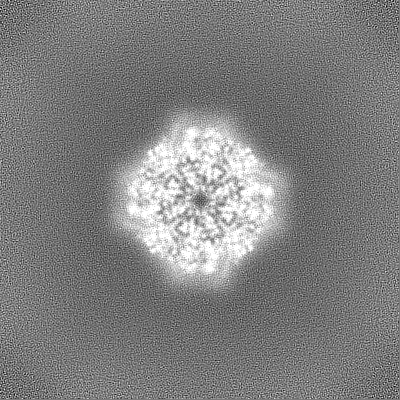
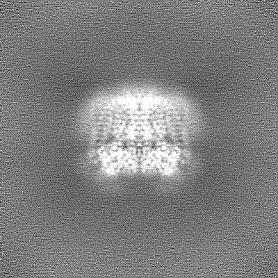
Journal: Proc Natl Acad Sci U S A / Year: 2025 Title: Pathogenic variants in the polycystin pore helix cause distinct forms of channel dysfunction. Authors: Orhi Esarte Palomero / Eduardo Guadarrama / Paul G DeCaen / Abstract: PKD2 is a member of the polycystin subfamily of transient receptor potential (TRP) ion channel subunits which traffic and function in primary cilia organelle membranes. Millions of individuals carry ...PKD2 is a member of the polycystin subfamily of transient receptor potential (TRP) ion channel subunits which traffic and function in primary cilia organelle membranes. Millions of individuals carry pathogenic genetic variants in PKD2 that cause a life-threatening condition called autosomal dominant polycystic kidney disease (ADPKD). Although ADPKD is a common monogenetic disorder, there is no drug cure or available therapeutics which address the underlying channel dysregulation. Furthermore, the structural and mechanistic impacts of most disease-causing variants are uncharacterized. Using direct cilia electrophysiology, cryogenic electron microscopy (cryo-EM), and superresolution imaging, we have found mechanistic differences in channel dysregulation caused by three germline missense variants located in PKD2's pore helix 1. Variant C632R reduces protein thermal stability, resulting in impaired channel assembly and abolishes primary cilia trafficking. In contrast, variants F629S and R638C retain native cilia trafficking but exhibit gating defects. Cryo-EM structures (2.7 to 2.8 Å resolution) indicate loss of critical pore helix interactions which precipitate allosteric collapse of the channels inner gate. Results demonstrate how ADPKD-causing mutations cause mechanistically divergent and ranging impacts on PKD2 function, despite their shared structural proximity. These unexpected findings highlight the need for structural and biophysical characterization of polycystin variants, which will guide rational drug development of ADPKD therapeutics.
Details: 25 mM HEPES-NaOH, 150 mM NaCl, 1 mM CaCl2, 1 mM TCEP
Grid
Model: Quantifoil R2/1 / Material: COPPER / Mesh: 300 / Support film - Material: CARBON / Support film - topology: HOLEY ARRAY / Pretreatment - Type: GLOW DISCHARGE / Pretreatment - Time: 20 sec. / Pretreatment - Atmosphere: AIR / Pretreatment - Pressure: 0.026000000000000002 kPa / Details: 15 mA
Vitrification
Cryogen name: ETHANE / Chamber humidity: 95 % / Chamber temperature: 278 K / Instrument: LEICA EM GP Details: Vitrification carried in air. Ethane temperature -183 C.
Details
Stabilized in amphipol A8-35. Monodisperse sample after gel filtration in a Superdex 200 column.
-
Electron microscopy
Microscope
TFS KRIOS
Specialist optics
Energy filter - Name: GIF Bioquantum / Energy filter - Slit width: 10 eV
Image recording
Film or detector model: GATAN K3 (6k x 4k) / Digitization - Dimensions - Width: 11520 pixel / Digitization - Dimensions - Height: 8184 pixel / Number grids imaged: 1 / Number real images: 9145 / Average electron dose: 60.0 e/Å2 / Details: 50 frame movie stacks. Super-resolution mode
Electron beam
Acceleration voltage: 300 kV / Electron source: FIELD EMISSION GUN
In the structure databanks used in Yorodumi, some data are registered as the other names, "COVID-19 virus" and "2019-nCoV". Here are the details of the virus and the list of structure data.
Jan 31, 2019. EMDB accession codes are about to change! (news from PDBe EMDB page)
EMDB accession codes are about to change! (news from PDBe EMDB page)
The allocation of 4 digits for EMDB accession codes will soon come to an end. Whilst these codes will remain in use, new EMDB accession codes will include an additional digit and will expand incrementally as the available range of codes is exhausted. The current 4-digit format prefixed with “EMD-” (i.e. EMD-XXXX) will advance to a 5-digit format (i.e. EMD-XXXXX), and so on. It is currently estimated that the 4-digit codes will be depleted around Spring 2019, at which point the 5-digit format will come into force.
The EM Navigator/Yorodumi systems omit the EMD- prefix.
Related info.:Q: What is EMD? / ID/Accession-code notation in Yorodumi/EM Navigator
Yorodumi is a browser for structure data from EMDB, PDB, SASBDB, etc.
This page is also the successor to EM Navigator detail page, and also detail information page/front-end page for Omokage search.
The word "yorodu" (or yorozu) is an old Japanese word meaning "ten thousand". "mi" (miru) is to see.
Related info.:EMDB / PDB / SASBDB / Comparison of 3 databanks / Yorodumi Search / Aug 31, 2016. New EM Navigator & Yorodumi / Yorodumi Papers / Jmol/JSmol / Function and homology information / Changes in new EM Navigator and Yorodumi
 Movie
Movie Controller


 Open data
 Basic information
 Map data
 Sample
 Keywords
 Function and homology information
 Homo sapiens (human)
 Authors
 United States, 5 items
  Citation
 Structure visualization
 Downloads & links
 emd_47260.png
 http://ftp.pdbj.org/pub/emdb/structures/EMD-47260


 Z (Sec.)
 Y (Row.)
 X (Col.)


 Sample components
 Processing
 Electron microscopy
 FIELD EMISSION GUN

