negative regulation of monoatomic ion transmembrane transport / positive regulation by virus of viral protein levels in host cell / spindle assembly involved in female meiosis / epigenetic programming in the zygotic pronuclei / UV-damage excision repair / biological process involved in interaction with symbiont / limb development / regulation of mitotic cell cycle phase transition / WD40-repeat domain binding / Cul4A-RING E3 ubiquitin ligase complex ...negative regulation of monoatomic ion transmembrane transport / positive regulation by virus of viral protein levels in host cell / spindle assembly involved in female meiosis / epigenetic programming in the zygotic pronuclei / UV-damage excision repair / biological process involved in interaction with symbiont / limb development / regulation of mitotic cell cycle phase transition / WD40-repeat domain binding / Cul4A-RING E3 ubiquitin ligase complex / Cul4-RING E3 ubiquitin ligase complex / Cul4B-RING E3 ubiquitin ligase complex / ubiquitin ligase complex scaffold activity / negative regulation of reproductive process / negative regulation of developmental process / locomotory exploration behavior / viral release from host cell / cullin family protein binding / ectopic germ cell programmed cell death / positive regulation of viral genome replication / positive regulation of Wnt signaling pathway / negative regulation of protein-containing complex assembly / proteasomal protein catabolic process / positive regulation of gluconeogenesis / nucleotide-excision repair / sperm end piece / positive regulation of protein-containing complex assembly / regulation of circadian rhythm / Recognition of DNA damage by PCNA-containing replication complex / DNA Damage Recognition in GG-NER / Dual Incision in GG-NER / Wnt signaling pathway / Transcription-Coupled Nucleotide Excision Repair (TC-NER) / Formation of TC-NER Pre-Incision Complex / Formation of Incision Complex in GG-NER / Dual incision in TC-NER / positive regulation of protein catabolic process / Gap-filling DNA repair synthesis and ligation in TC-NER / cellular response to UV / rhythmic process / site of double-strand break / sperm principal piece / Neddylation / sperm midpiece / Potential therapeutics for SARS / ubiquitin-dependent protein catabolic process / damaged DNA binding / transmembrane transporter binding / proteasome-mediated ubiquitin-dependent protein catabolic process / protein-macromolecule adaptor activity / chromosome, telomeric region / protein ubiquitination / DNA repair / apoptotic process / DNA damage response / negative regulation of apoptotic process / protein-containing complex binding / nucleolus / perinuclear region of cytoplasm / protein-containing complex / : / DNA binding / extracellular exosome / nucleoplasm / membrane / metal ion binding / nucleus / cytoplasm / cytosol Similarity search - Function
Yippee/Mis18/Cereblon / Yippee zinc-binding/DNA-binding /Mis18, centromere assembly / CULT domain / CULT domain profile. / Lon N-terminal domain profile. / Lon protease, N-terminal domain / Lon protease, N-terminal domain superfamily / ATP-dependent protease La (LON) substrate-binding domain / Found in ATP-dependent protease La (LON) / : ...Yippee/Mis18/Cereblon / Yippee zinc-binding/DNA-binding /Mis18, centromere assembly / CULT domain / CULT domain profile. / Lon N-terminal domain profile. / Lon protease, N-terminal domain / Lon protease, N-terminal domain superfamily / ATP-dependent protease La (LON) substrate-binding domain / Found in ATP-dependent protease La (LON) / : / RSE1/DDB1/CPSF1 second beta-propeller / Cleavage/polyadenylation specificity factor, A subunit, C-terminal / Cleavage/polyadenylation specificity factor, A subunit, N-terminal / : / CPSF A subunit region / RSE1/DDB1/CPSF1 first beta-propeller / PUA-like superfamily / WD40-repeat-containing domain superfamily / WD40/YVTN repeat-like-containing domain superfamily Similarity search - Domain/homology
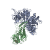
Journal: ACS Med Chem Lett / Year: 2025 Title: Development of a Buchwald-Hartwig Amination for an Accelerated Library Synthesis of Cereblon Binders. Authors: Anastasia Lejava / Giulianna A Miseo / Thomas Phan / Jinyi Zhu / Hannah L Powers / Jianqing Li / Deborah S Mortensen / Christoph W Zapf / Gody Khambatta / Jennifer Buenviaje / Natalie Holmberg-Douglas / Abstract: In recent years, targeted protein degradation (TPD) has emerged as a powerful therapeutic modality utilizing both heterobifunctional ligand-directed degraders (LDDs) and molecular glues (e.g., ...In recent years, targeted protein degradation (TPD) has emerged as a powerful therapeutic modality utilizing both heterobifunctional ligand-directed degraders (LDDs) and molecular glues (e.g., CELMoDs) to recruit E3 ligases for inducing polyubiquitination and subsequent proteasomal degradation of target proteins. The immunomodulatory drugs lenalidomide and pomalidomide bind to cereblon (CRBN), a substrate receptor of the CRL4A E3 ligase complex, to initiate degradation of neosubstrates critical for cell survival. Recently, nonlenalidomide or pomalidomide CRBN binders, known as alternate glutarimides, have gained popularity, offering potential degraders with varying physicochemical properties. Specifically, 3-substituted indazole derivatives have emerged as potent CRBN binders. We developed conditions for the direct cross-coupling of unprotected glutarimides with amines, streamlining the synthesis of alternative CRBN binders. This manuscript describes the rapid synthesis of 30 CRBN binders, their characterization as potential degraders and a cryo-EM structure of the CRBN/DDB1 with a representative compound ().
Cryogen name: ETHANE / Chamber humidity: 100 % / Chamber temperature: 278 K / Instrument: FEI VITROBOT MARK IV
-
Electron microscopy
Microscope
TFS KRIOS
Image recording
#0 - Image recording ID: 1 / #0 - Film or detector model: FEI FALCON IV (4k x 4k) / #0 - Number grids imaged: 1 / #0 - Number real images: 11921 / #0 - Average exposure time: 3.03 sec. / #0 - Average electron dose: 27.67 e/Å2 / #1 - Image recording ID: 2 / #1 - Film or detector model: FEI FALCON IV (4k x 4k) / #1 - Number grids imaged: 1 / #1 - Number real images: 8200 / #1 - Average exposure time: 3.49 sec. / #1 - Average electron dose: 39.28 e/Å2 / #1 - Details: 30 degree tilt
Electron beam
Acceleration voltage: 300 kV / Electron source: FIELD EMISSION GUN
In the structure databanks used in Yorodumi, some data are registered as the other names, "COVID-19 virus" and "2019-nCoV". Here are the details of the virus and the list of structure data.
Jan 31, 2019. EMDB accession codes are about to change! (news from PDBe EMDB page)
EMDB accession codes are about to change! (news from PDBe EMDB page)
The allocation of 4 digits for EMDB accession codes will soon come to an end. Whilst these codes will remain in use, new EMDB accession codes will include an additional digit and will expand incrementally as the available range of codes is exhausted. The current 4-digit format prefixed with “EMD-” (i.e. EMD-XXXX) will advance to a 5-digit format (i.e. EMD-XXXXX), and so on. It is currently estimated that the 4-digit codes will be depleted around Spring 2019, at which point the 5-digit format will come into force.
The EM Navigator/Yorodumi systems omit the EMD- prefix.
Related info.:Q: What is EMD? / ID/Accession-code notation in Yorodumi/EM Navigator
Yorodumi is a browser for structure data from EMDB, PDB, SASBDB, etc.
This page is also the successor to EM Navigator detail page, and also detail information page/front-end page for Omokage search.
The word "yorodu" (or yorozu) is an old Japanese word meaning "ten thousand". "mi" (miru) is to see.
Related info.:EMDB / PDB / SASBDB / Comparison of 3 databanks / Yorodumi Search / Aug 31, 2016. New EM Navigator & Yorodumi / Yorodumi Papers / Jmol/JSmol / Function and homology information / Changes in new EM Navigator and Yorodumi
 Movie
Movie Controller

 Yorodumi
 Open data
 Basic information
 Map data
 Sample
 Keywords
 Function and homology information
 Homo sapiens (human)
 Authors
 Citation
 Structure visualization
 Downloads & links
 emd_47111.png
 http://ftp.pdbj.org/pub/emdb/structures/EMD-47111


 Z (Sec.)
 Y (Row.)
 X (Col.)


 Sample components
 Spodoptera frugiperda (fall armyworm)
 Processing
 Electron microscopy
 FIELD EMISSION GUN