Movie
Movie Controller
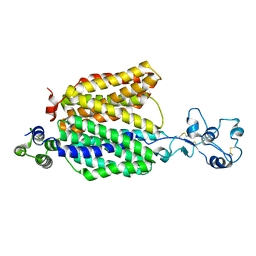


+ Open data

- Basic information
| Entry |  | |||||||||
|---|---|---|---|---|---|---|---|---|---|---|
| Title | Structure of URAT1 in complex with TD-3 | |||||||||
 Map data Map data | full map | |||||||||
 Sample Sample |
| |||||||||
 Keywords Keywords | Membrane protein / membrane transporter / TRANSPORT PROTEIN | |||||||||
| Biological species |  Homo sapiens (human) Homo sapiens (human) | |||||||||
| Method | single particle reconstruction / cryo EM / Resolution: 2.55 Å | |||||||||
 Authors Authors | Suo Y / Fedor JG / Lee S-Y | |||||||||
| Funding support |  United States, 1 items United States, 1 items
| |||||||||
 Citation Citation | Journal: Nat Commun / Year: 2025 Title: Molecular basis of the urate transporter URAT1 inhibition by gout drugs. Authors: Suo Y / Fedor JG / Zhang H / Tsolova K / Shi X / Sharma K / Kumari S / Borgnia M / Zhan P / Im W / Lee SY | |||||||||
| History |
|
- Structure visualization
- Downloads & links
-EMDB archive
| Map data | emd_46951.map.gz | 59.8 MB |  EMDB map data format EMDB map data format | |
|---|---|---|---|---|
| Header (meta data) | emd-46951-v30.xmlemd-46951.xml | 19.6 KB 19.6 KB | Display Display | EMDB header |
| Images |  emd_46951.png emd_46951.png | 89.9 KB | ||
| Filedesc metadata | emd-46951.cif.gz | 6.6 KB | ||
| Others | emd_46951_half_map_1.map.gzemd_46951_half_map_2.map.gz | 59.5 MB 59.5 MB | ||
| Archive directory |  http://ftp.pdbj.org/pub/emdb/structures/EMD-46951ftp://ftp.pdbj.org/pub/emdb/structures/EMD-46951 http://ftp.pdbj.org/pub/emdb/structures/EMD-46951ftp://ftp.pdbj.org/pub/emdb/structures/EMD-46951 | HTTPS FTP |
-Validation report
| Summary document | emd_46951_validation.pdf.gz | 810.4 KB | Display | EMDB validaton report |
|---|---|---|---|---|
| Full document | emd_46951_full_validation.pdf.gz | 810 KB | Display | |
| Data in XML | emd_46951_validation.xml.gz | 12.3 KB | Display | |
| Data in CIF | emd_46951_validation.cif.gz | 14.4 KB | Display | |
| Arichive directory | https://ftp.pdbj.org/pub/emdb/validation_reports/EMD-46951ftp://ftp.pdbj.org/pub/emdb/validation_reports/EMD-46951 | HTTPS FTP |
-Related structure data
| Related structure data |  9dkcMC  9dk9C  9dkaC  9dkbC M: atomic model generated by this map C: citing same article ( |
|---|


-Links
| EMDB pages | EMDB (EBI/PDBe) / EMDataResource |
|---|
-Map
| File | Download / File: emd_46951.map.gz / Format: CCP4 / Size: 64 MB / Type: IMAGE STORED AS FLOATING POINT NUMBER (4 BYTES) | ||||||||||||||||||||
|---|---|---|---|---|---|---|---|---|---|---|---|---|---|---|---|---|---|---|---|---|---|
| Annotation | full map | ||||||||||||||||||||
| Voxel size | X=Y=Z: 0.8469 Å | ||||||||||||||||||||
| Density |
| ||||||||||||||||||||
| Symmetry | Space group: 1 | ||||||||||||||||||||
| Details | EMDB XML:
|
-Supplemental data
- Sample components
-Entire : URAT1
| Entire | Name: URAT1 |
|---|---|
| Components |
|
-Supramolecule #1: URAT1
| Supramolecule | Name: URAT1 / type: complex / ID: 1 / Parent: 0 / Macromolecule list: #1 |
|---|---|
| Source (natural) | Organism: Homo sapiens (human) |
| Molecular weight | Theoretical: 55 KDa |
-Macromolecule #1: URAT1
| Macromolecule | Name: URAT1 / type: protein_or_peptide / ID: 1 / Number of copies: 1 / Enantiomer: LEVO |
|---|---|
| Source (natural) | Organism: Homo sapiens (human) |
| Molecular weight | Theoretical: 55.379844 KDa |
| Recombinant expression | Organism: Homo sapiens (human) |
| Sequence | String: MAFSELLDQV GGLGRFQVLQ TVALVVPIMW LCTQSMLENF SAAVPSHRCW VPLLDNSTAQ ASVPGALGPE ALLAVSIPPG PNQGPHQCR RFRQPQWQLL DPNATATNWS EAATEPCVDG WVYDRSTFTS TIVAKWDLVC DSQALKPMAQ SIYLAGILVG A AVCGPASD ...String: MAFSELLDQV GGLGRFQVLQ TVALVVPIMW LCTQSMLENF SAAVPSHRCW VPLLDNSTAQ ASVPGALGPE ALLAVSIPPG PNQGPHQCR RFRQPQWQLL DPNATATNWS EAATEPCVDG WVYDRSTFTS TIVAKWDLVC DSQALKPMAQ SIYLAGILVG A AVCGPASD RFGRRLVLTW SYLQMAVSGT AAAFAPTFPV YCLFRFLVAF AVAGVMMNTG TLVMEWTSAQ ARPLVMTLNS LG FSFGHVL MAAVAYGVRD WALLQLVVSV PFFLCFVYSC WLAESARWLL ITGRLDRGLR ELQRVAAING KRAVGDTLTP QVL LSAMQE ELSVGQAPAS LGTLLRTPGL RLRTCISTLC WFAFGFTFFG LALDLQALGS NIFLLQVLIG VVDIPAKIGT LLLL SRLGR RPTQAASLVL AGLCILANTL VPHEMGALRS ALAVLGLGGL GAAFTCITIY SGELFPTVLR MTAVGLGQMA ARGGA ILGP LVRLLGVHGP WLPLLVYGTV PVLSGLAALL LPET |
-Macromolecule #2: 2-({1-[(4-bromonaphthalen-1-yl)methyl]-1H-imidazo[4,5-b]pyridin-2...
| Macromolecule | Name: 2-({1-[(4-bromonaphthalen-1-yl)methyl]-1H-imidazo[4,5-b]pyridin-2-yl}sulfanyl)-2-methylpropanoic acid type: ligand / ID: 2 / Number of copies: 1 / Formula: A1A45 |
|---|---|
| Molecular weight | Theoretical: 456.356 Da |
-Experimental details
-Structure determination
| Method | cryo EM |
|---|---|
 Processing Processing | single particle reconstruction |
| Aggregation state | particle |
-Sample preparation
| Concentration | 10 mg/mL | ||||||||||||
|---|---|---|---|---|---|---|---|---|---|---|---|---|---|
| Buffer | pH: 8 Component:
| ||||||||||||
| Grid | Model: Quantifoil R1.2/1.3 / Support film - Material: GOLD / Support film - topology: HOLEY / Pretreatment - Type: GLOW DISCHARGE / Pretreatment - Time: 300 sec. / Pretreatment - Atmosphere: AIR / Pretreatment - Pressure: 0.00039000000000000005 kPa | ||||||||||||
| Vitrification | Cryogen name: ETHANE / Chamber humidity: 95 % / Chamber temperature: 280 K / Instrument: LEICA EM GP | ||||||||||||
| Details | Monodisperse sample |
- Electron microscopy
| Microscope | TFS KRIOS |
|---|---|
| Temperature | Max: 70.0 K |
| Specialist optics | Energy filter - Name: GIF Bioquantum / Energy filter - Slit width: 20 eV |
| Image recording | Film or detector model: GATAN K3 (6k x 4k) / Digitization - Dimensions - Width: 5760 pixel / Digitization - Dimensions - Height: 4092 pixel / Number grids imaged: 1 / Number real images: 18880 / Average exposure time: 1.8 sec. / Average electron dose: 50.0 e/Å2 |
| Electron beam | Acceleration voltage: 300 kV / Electron source:  FIELD EMISSION GUN FIELD EMISSION GUN |
| Electron optics | C2 aperture diameter: 100.0 µm / Illumination mode: FLOOD BEAM / Imaging mode: BRIGHT FIELD / Cs: 2.7 mm / Nominal defocus max: 2.0 µm / Nominal defocus min: 1.0 µm / Nominal magnification: 105000 |
| Sample stage | Specimen holder model: FEI TITAN KRIOS AUTOGRID HOLDER / Cooling holder cryogen: NITROGEN |
| Experimental equipment |  Model: Titan Krios / Image courtesy: FEI Company |