National Institutes of Health/National Institute of General Medical Sciences (NIH/NIGMS)
GM130234
United States
Citation
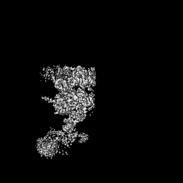
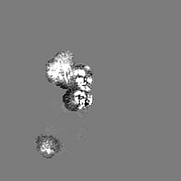
Journal: J Cell Biol / Year: 2025 Title: Structural insights into the coupling between VCP, an essential unfoldase, and a deubiquitinase. Authors: Lauren E Vostal / Noa E Dahan / Matthew J Reynolds / Lily I Kronenberg / Tarun M Kapoor / Abstract: Proteostasis involves degradation and recycling of proteins from organelles, membranes, and multiprotein complexes. These processes can depend on protein extraction and unfolding by the essential ...Proteostasis involves degradation and recycling of proteins from organelles, membranes, and multiprotein complexes. These processes can depend on protein extraction and unfolding by the essential mechanoenzyme valosin-containing protein (VCP) and on ubiquitin chain remodeling by ubiquitin-specific proteases known as deubiquitinases (DUBs). How the activities of VCP and DUBs are coordinated is poorly understood. Here, we focus on the DUB VCPIP1, a VCP interactor required for post-mitotic Golgi and ER organization. We determine ∼3.3 Å cryogenic electron microscopy structures of VCP-VCPIP1 complexes in the absence of added nucleotide or the presence of an ATP analog. We find that up to 3 VCPIP1 protomers interact with the VCP hexamer to position VCPIP1's catalytic domain at the exit of VCP's central pore, poised to cleave ubiquitin following substrate unfolding. We observe competition between VCPIP1 and other cofactors for VCP binding and show that VCP stimulates VCPIP1's DUB activity. Together, our data suggest how the two enzyme activities can be coordinated to regulate proteostasis.
In the structure databanks used in Yorodumi, some data are registered as the other names, "COVID-19 virus" and "2019-nCoV". Here are the details of the virus and the list of structure data.
Jan 31, 2019. EMDB accession codes are about to change! (news from PDBe EMDB page)
EMDB accession codes are about to change! (news from PDBe EMDB page)
The allocation of 4 digits for EMDB accession codes will soon come to an end. Whilst these codes will remain in use, new EMDB accession codes will include an additional digit and will expand incrementally as the available range of codes is exhausted. The current 4-digit format prefixed with “EMD-” (i.e. EMD-XXXX) will advance to a 5-digit format (i.e. EMD-XXXXX), and so on. It is currently estimated that the 4-digit codes will be depleted around Spring 2019, at which point the 5-digit format will come into force.
The EM Navigator/Yorodumi systems omit the EMD- prefix.
Related info.:Q: What is EMD? / ID/Accession-code notation in Yorodumi/EM Navigator
Yorodumi is a browser for structure data from EMDB, PDB, SASBDB, etc.
This page is also the successor to EM Navigator detail page, and also detail information page/front-end page for Omokage search.
The word "yorodu" (or yorozu) is an old Japanese word meaning "ten thousand". "mi" (miru) is to see.
Related info.:EMDB / PDB / SASBDB / Comparison of 3 databanks / Yorodumi Search / Aug 31, 2016. New EM Navigator & Yorodumi / Yorodumi Papers / Jmol/JSmol / Function and homology information / Changes in new EM Navigator and Yorodumi
 Movie
Movie Controller


 Open data
 Basic information
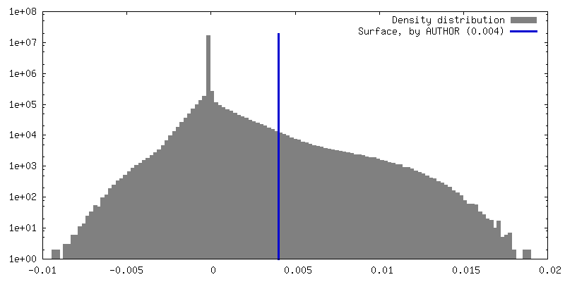
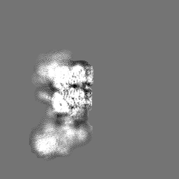
 Map data
 Sample
 Keywords
 Function and homology information
 Homo sapiens (human)
 Authors
 United States, 1 items
  Citation
 Structure visualization
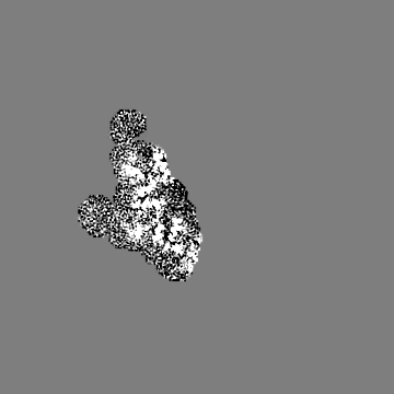
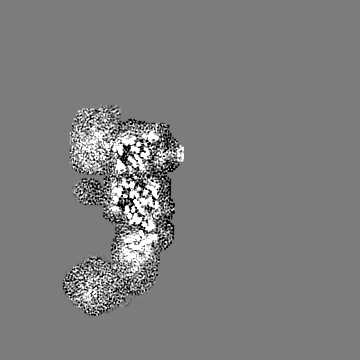
 Downloads & links
 emd_46912.png
 http://ftp.pdbj.org/pub/emdb/structures/EMD-46912


 Z (Sec.)
 Y (Row.)
 X (Col.)


 Sample components

 Processing
 Electron microscopy
 FIELD EMISSION GUN

