National Institutes of Health/National Institute of General Medical Sciences (NIH/NIGMS)
P41GM136508
United States
Department of Defense (DOD, United States)
HDTRA1-21-1-0004
United States
Howard Hughes Medical Institute (HHMI)
United States
Citation
Journal: bioRxiv / Year: 2024 Title: Energy filtering enables macromolecular MicroED data at sub-atomic resolution. Authors: Max T B Clabbers / Johan Hattne / Michael W Martynowycz / Tamir Gonen / Abstract: High resolution information is important for accurate structure modelling. However, this level of detail is typically difficult to attain in macromolecular crystallography because the diffracted ...High resolution information is important for accurate structure modelling. However, this level of detail is typically difficult to attain in macromolecular crystallography because the diffracted intensities rapidly fade with increasing resolution. The problem cannot be circumvented by increasing the fluence as this leads to detrimental radiation damage. Previously, we demonstrated that high quality MicroED data can be obtained at low flux conditions using electron counting with direct electron detectors. The improved sensitivity and accuracy of these detectors essentially eliminate the read-out noise, such that the measurement of faint high-resolution reflections is limited by other sources of noise. Inelastic scattering is a major contributor of such noise, increasing background counts and broadening diffraction spots. Here, we demonstrate that a substantial improvement in signal-to-noise ratio can be achieved using an energy filter to largely remove the inelastically scattered electrons. This strategy resulted in sub-atomic resolution MicroED data from proteinase K crystals, enabling accurate structure modelling and the visualization of detailed features. Interestingly, filtering out the noise revealed diffuse scattering phenomena that can hold additional structural information. Our findings suggest that combining energy filtering and electron counting can provide more accurate measurements at higher resolution, providing better insights into protein function and facilitating more precise model refinement.
Model: Quantifoil R2/2 / Material: COPPER / Mesh: 200 / Support film - Material: CARBON / Support film - topology: HOLEY / Support film - Film thickness: 10 / Pretreatment - Type: GLOW DISCHARGE / Pretreatment - Time: 60 sec. / Details: Negative 15 mA
Energy filter - Name: TFS Selectris / Energy filter - Slit width: 10 eV
Image recording
Film or detector model: FEI FALCON IV (4k x 4k) / Digitization - Dimensions - Width: 4096 pixel / Digitization - Dimensions - Height: 4096 pixel / Number grids imaged: 1 / Number real images: 1 / Number diffraction images: 420 / Average exposure time: 1.0 sec. / Average electron dose: 0.002 e/Å2
Electron beam
Acceleration voltage: 300 kV / Electron source: FIELD EMISSION GUN
In the structure databanks used in Yorodumi, some data are registered as the other names, "COVID-19 virus" and "2019-nCoV". Here are the details of the virus and the list of structure data.
Jan 31, 2019. EMDB accession codes are about to change! (news from PDBe EMDB page)
EMDB accession codes are about to change! (news from PDBe EMDB page)
The allocation of 4 digits for EMDB accession codes will soon come to an end. Whilst these codes will remain in use, new EMDB accession codes will include an additional digit and will expand incrementally as the available range of codes is exhausted. The current 4-digit format prefixed with “EMD-” (i.e. EMD-XXXX) will advance to a 5-digit format (i.e. EMD-XXXXX), and so on. It is currently estimated that the 4-digit codes will be depleted around Spring 2019, at which point the 5-digit format will come into force.
The EM Navigator/Yorodumi systems omit the EMD- prefix.
Related info.:Q: What is EMD? / ID/Accession-code notation in Yorodumi/EM Navigator
Yorodumi is a browser for structure data from EMDB, PDB, SASBDB, etc.
This page is also the successor to EM Navigator detail page, and also detail information page/front-end page for Omokage search.
The word "yorodu" (or yorozu) is an old Japanese word meaning "ten thousand". "mi" (miru) is to see.
Related info.:EMDB / PDB / SASBDB / Comparison of 3 databanks / Yorodumi Search / Aug 31, 2016. New EM Navigator & Yorodumi / Yorodumi Papers / Jmol/JSmol / Function and homology information / Changes in new EM Navigator and Yorodumi
 Movie
Movie Controller


 Open data
 Basic information
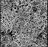
 Map data
 Sample
 Keywords
 Function and homology information
 Parengyodontium album (fungus)
 Authors
 United States, 3 items
  Citation
 Structure visualization
 Downloads & links
 emd_46871.jpg
 http://ftp.pdbj.org/pub/emdb/structures/EMD-46871
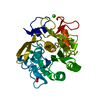


 X (Sec.)
 Y (Row.)
 Z (Col.)


 Sample components

 Processing
 Electron microscopy
 FIELD EMISSION GUN

