Movie
Movie Controller


+ Open data

- Basic information
| Entry |  | |||||||||
|---|---|---|---|---|---|---|---|---|---|---|
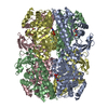
| Title | Focused region on azoRhuA-bCDRhuA co-assembled nanotubes | |||||||||
 Map data Map data | ||||||||||
 Sample Sample |
| |||||||||
 Keywords Keywords | Assembly / LYASE | |||||||||
| Function / homology |  Function and homology information Function and homology informationrhamnulose-1-phosphate aldolase / rhamnulose-1-phosphate aldolase activity / rhamnose catabolic process / pentose catabolic process / aldehyde-lyase activity / metal ion binding / identical protein binding / cytosol Similarity search - Function | |||||||||
| Biological species |  | |||||||||
| Method | single particle reconstruction / cryo EM / Resolution: 3.03 Å | |||||||||
 Authors Authors | Zhang Z / Sonani RR / Wang F / Egelman EH / Tezcan FA | |||||||||
| Funding support |  United States, 1 items United States, 1 items
| |||||||||
 Citation Citation | Journal: Chem / Year: 2025 Title: Design of light- and chemically responsive protein assemblies through host-guest interactions. Authors: Zhiyin Zhang / Huat T Chiang / Ying Xia / Nicole Avakyan / Ravi R Sonani / Fengbin Wang / Edward H Egelman / James J De Yoreo / Lilo D Pozzo / F Akif Tezcan / Abstract: Host-guest interactions have been widely used to build responsive materials and molecular machines owing to their inherently dynamic nature, interaction specificity, and responsiveness to diverse ...Host-guest interactions have been widely used to build responsive materials and molecular machines owing to their inherently dynamic nature, interaction specificity, and responsiveness to diverse stimuli. Here we have set out to exploit these advantages of host-guest chemistry in the design of dynamic protein assemblies, using a symmetric protein, RhuA, as a building block. We show that RhuA variant individually modified with β-cyclodextrin (βCD) (host) or azobenzene (guest) functionalities can specifically pair with each other to form highly ordered 1- and 2-D assemblies. Association and dissociation RhuA-RhuA assemblies can be controlled by UV and visible light as well as by small-molecule modulators of βCD-azobenzene interactions. Kinetics analyses reveal that RhuA-RhuA nanotubes assemble without a nucleation barrier, a highly unusual occurrence for helical supramolecular systems. Taken together, our findings provide a compelling example for achieving complex structural and dynamic outcomes in protein assembly through simple chemical design. | |||||||||
| History |
|
- Structure visualization
| Supplemental images |
|---|

- Downloads & links
-EMDB archive
| Map data | emd_46826.map.gz | 203.4 MB | EMDB map data format | |
|---|---|---|---|---|
| Header (meta data) | emd-46826-v30.xmlemd-46826.xml | 20.9 KB 20.9 KB | Display Display | EMDB header |
| FSC (resolution estimation) | emd_46826_fsc.xml | 12.7 KB | Display | FSC data file |
| Images |  emd_46826.png emd_46826.png | 121.6 KB | ||
| Filedesc metadata | emd-46826.cif.gz | 6.2 KB | ||
| Others | emd_46826_additional_1.map.gzemd_46826_additional_2.map.gzemd_46826_half_map_1.map.gzemd_46826_half_map_2.map.gz | 30.8 MB 931.6 MB 198.4 MB 198.4 MB | ||
| Archive directory |  http://ftp.pdbj.org/pub/emdb/structures/EMD-46826ftp://ftp.pdbj.org/pub/emdb/structures/EMD-46826 http://ftp.pdbj.org/pub/emdb/structures/EMD-46826ftp://ftp.pdbj.org/pub/emdb/structures/EMD-46826 | HTTPS FTP |
-Related structure data
| Related structure data |  9dghMC M: atomic model generated by this map C: citing same article ( |
|---|---|
| Similar structure data |


-Links
| EMDB pages | EMDB (EBI/PDBe) / EMDataResource |
|---|
-Map
| File | Download / File: emd_46826.map.gz / Format: CCP4 / Size: 216 MB / Type: IMAGE STORED AS FLOATING POINT NUMBER (4 BYTES) | ||||||||||||||||||||||||||||||||||||
|---|---|---|---|---|---|---|---|---|---|---|---|---|---|---|---|---|---|---|---|---|---|---|---|---|---|---|---|---|---|---|---|---|---|---|---|---|---|
| Projections & slices | Image control
Images are generated by Spider. | ||||||||||||||||||||||||||||||||||||
| Voxel size | X=Y=Z: 0.86 Å | ||||||||||||||||||||||||||||||||||||
| Density |
| ||||||||||||||||||||||||||||||||||||
| Symmetry | Space group: 1 | ||||||||||||||||||||||||||||||||||||
| Details | EMDB XML:
|
 Z (Sec.)
 Y (Row.)
 X (Col.)


-Supplemental data
-Additional map: local filtered volume map
| File | emd_46826_additional_1.map | ||||||||||||
|---|---|---|---|---|---|---|---|---|---|---|---|---|---|
| Annotation | local filtered volume map | ||||||||||||
| Projections & Slices |
| ||||||||||||
| Density Histograms |


-Additional map: EMready processed volume map
| File | emd_46826_additional_2.map | ||||||||||||
|---|---|---|---|---|---|---|---|---|---|---|---|---|---|
| Annotation | EMready processed volume map | ||||||||||||
| Projections & Slices |
| ||||||||||||
| Density Histograms |


-Half map: #1
| File | emd_46826_half_map_1.map | ||||||||||||
|---|---|---|---|---|---|---|---|---|---|---|---|---|---|
| Projections & Slices |
| ||||||||||||
| Density Histograms |


-Half map: #2
| File | emd_46826_half_map_2.map | ||||||||||||
|---|---|---|---|---|---|---|---|---|---|---|---|---|---|
| Projections & Slices |
| ||||||||||||
| Density Histograms |


- Sample components
-Entire : Helical assembly of azobenzene and beta-cyclodextrin modified RhuA
| Entire | Name: Helical assembly of azobenzene and beta-cyclodextrin modified RhuA |
|---|---|
| Components |
|
-Supramolecule #1: Helical assembly of azobenzene and beta-cyclodextrin modified RhuA
| Supramolecule | Name: Helical assembly of azobenzene and beta-cyclodextrin modified RhuA type: complex / ID: 1 / Parent: 0 / Macromolecule list: #1 |
|---|---|
| Source (natural) | Organism: |
-Macromolecule #1: Rhamnulose-1-phosphate aldolase
| Macromolecule | Name: Rhamnulose-1-phosphate aldolase / type: protein_or_peptide / ID: 1 / Number of copies: 16 / Enantiomer: LEVO / EC number: rhamnulose-1-phosphate aldolase |
|---|---|
| Source (natural) | Organism: |
| Molecular weight | Theoretical: 30.088361 KDa |
| Recombinant expression | Organism: |
| Sequence | String: MQNITQSWFV QGMIKATTDA WLKGWDERNG GNLTLRLDDA DIAPYHDNFH QQPRYIPLSQ PMPLLANTPF IVTGSGKFFR NVQLDPAAN LGIVKVDSCG AGYHILWGLT NEAVPTSELP AHFLSHSERI KATNGKDRVI MHCHATNLIA LTYVLENDTA V FTRQLWEG ...String: MQNITQSWFV QGMIKATTDA WLKGWDERNG GNLTLRLDDA DIAPYHDNFH QQPRYIPLSQ PMPLLANTPF IVTGSGKFFR NVQLDPAAN LGIVKVDSCG AGYHILWGLT NEAVPTSELP AHFLSHSERI KATNGKDRVI MHCHATNLIA LTYVLENDTA V FTRQLWEG STECLVVFPD GVGILPWMVP GTDAIGQATA QEMQKHSLVL WPFHGVFGSG PTLDETFGLI DTAEKSAQVL VK VYSMGGM KQTISREELI ALGKRFGVTP LASALAL UniProtKB: Rhamnulose-1-phosphate aldolase |
-Macromolecule #3: ZINC ION
| Macromolecule | Name: ZINC ION / type: ligand / ID: 3 / Number of copies: 16 / Formula: ZN |
|---|---|
| Molecular weight | Theoretical: 65.409 Da |
-Macromolecule #4: 1-[4-(2-phenylhydrazin-1-yl)phenyl]pyrrolidine-2,5-dione
| Macromolecule | Name: 1-[4-(2-phenylhydrazin-1-yl)phenyl]pyrrolidine-2,5-dione type: ligand / ID: 4 / Number of copies: 2 / Formula: A1A4G |
|---|---|
| Molecular weight | Theoretical: 281.309 Da |
-Experimental details
-Structure determination
| Method | cryo EM |
|---|---|
 Processing Processing | single particle reconstruction |
| Aggregation state | helical array |
-Sample preparation
| Buffer | pH: 7.5 |
|---|---|
| Vitrification | Cryogen name: ETHANE |
- Electron microscopy
| Microscope | TFS KRIOS |
|---|---|
| Image recording | Film or detector model: GATAN K3 (6k x 4k) / Average electron dose: 50.0 e/Å2 |
| Electron beam | Acceleration voltage: 300 kV / Electron source:  FIELD EMISSION GUN FIELD EMISSION GUN |
| Electron optics | Illumination mode: FLOOD BEAM / Imaging mode: BRIGHT FIELD / Nominal defocus max: 2.1 µm / Nominal defocus min: 1.5 µm |
| Experimental equipment |  Model: Titan Krios / Image courtesy: FEI Company |