ムービー
ムービー コントローラー


+ データを開く

- 基本情報
基本情報
| 登録情報 | データベース: EMDB / ID: EMD-4672 | |||||||||
|---|---|---|---|---|---|---|---|---|---|---|
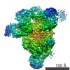
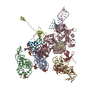
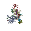
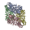
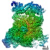
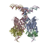
| タイトル | Human U5.U4/U6 tri-snRNP, multi-body refinement, Brr2 map | |||||||||
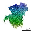
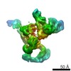
 マップデータ マップデータ | Human U5.U4/U6 tri-snRNP, multi-body refinement, Brr2 map | |||||||||
 試料 試料 |
| |||||||||
| 生物種 |  Homo sapiens (ヒト) Homo sapiens (ヒト) | |||||||||
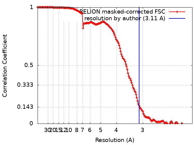
| 手法 | 単粒子再構成法 / クライオ電子顕微鏡法 / 解像度: 3.11 Å | |||||||||
 データ登録者 データ登録者 | Charenton C / Wilkinson ME / Nagai K | |||||||||
 引用 引用 | ジャーナル: Science / 年: 2019 タイトル: Mechanism of 5' splice site transfer for human spliceosome activation. 著者: Clément Charenton / Max E Wilkinson / Kiyoshi Nagai /  要旨: The prespliceosome, comprising U1 and U2 small nuclear ribonucleoproteins (snRNPs) bound to the precursor messenger RNA 5' splice site (5'SS) and branch point sequence, associates with the U4/U6.U5 ...The prespliceosome, comprising U1 and U2 small nuclear ribonucleoproteins (snRNPs) bound to the precursor messenger RNA 5' splice site (5'SS) and branch point sequence, associates with the U4/U6.U5 tri-snRNP to form the fully assembled precatalytic pre-B spliceosome. Here, we report cryo-electron microscopy structures of the human pre-B complex captured before U1 snRNP dissociation at 3.3-angstrom core resolution and the human tri-snRNP at 2.9-angstrom resolution. U1 snRNP inserts the 5'SS-U1 snRNA helix between the two RecA domains of the Prp28 DEAD-box helicase. Adenosine 5'-triphosphate-dependent closure of the Prp28 RecA domains releases the 5'SS to pair with the nearby U6 ACAGAGA-box sequence presented as a mobile loop. The structures suggest that formation of the 5'SS-ACAGAGA helix triggers remodeling of an intricate protein-RNA network to induce Brr2 helicase relocation to its loading sequence in U4 snRNA, enabling Brr2 to unwind the U4/U6 snRNA duplex to allow U6 snRNA to form the catalytic center of the spliceosome. | |||||||||
| 履歴 |
|
- 構造の表示
構造の表示
| ムービー |
 ムービービューア ムービービューア |
|---|---|
| 構造ビューア | EMマップ: SurfViewMolmilJmol/JSmol |
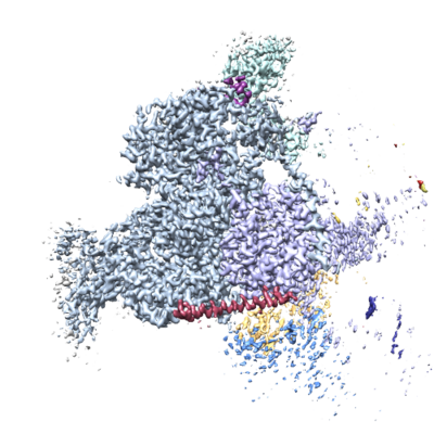
| 添付画像 |
- ダウンロードとリンク
-EMDBアーカイブ
| マップデータ | emd_4672.map.gz | 262.8 MB | EMDBマップデータ形式 | |
|---|---|---|---|---|
| ヘッダ (付随情報) | emd-4672-v30.xmlemd-4672.xml | 15.3 KB 15.3 KB | 表示 表示 | EMDBヘッダ |
| FSC (解像度算出) | emd_4672_fsc.xml | 14.8 KB | 表示 | FSCデータファイル |
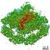
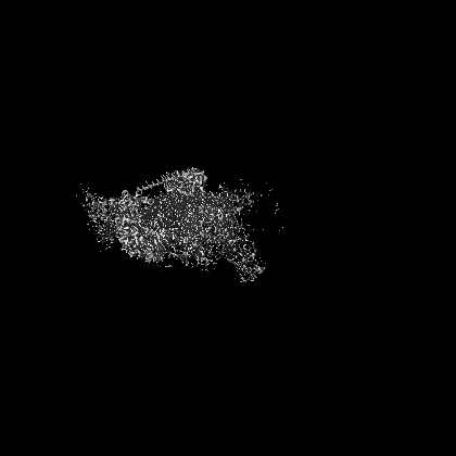
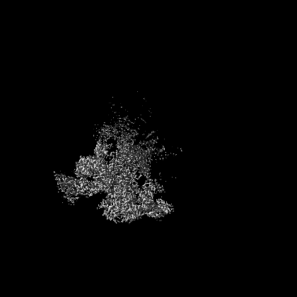
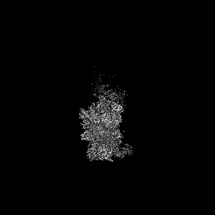
| 画像 |  emd_4672.png emd_4672.png | 207.2 KB | ||
| アーカイブディレクトリ |  http://ftp.pdbj.org/pub/emdb/structures/EMD-4672ftp://ftp.pdbj.org/pub/emdb/structures/EMD-4672 http://ftp.pdbj.org/pub/emdb/structures/EMD-4672ftp://ftp.pdbj.org/pub/emdb/structures/EMD-4672 | HTTPS FTP |
-検証レポート
| 文書・要旨 | emd_4672_validation.pdf.gz | 271.1 KB | 表示 | EMDB検証レポート |
|---|---|---|---|---|
| 文書・詳細版 | emd_4672_full_validation.pdf.gz | 270.2 KB | 表示 | |
| XML形式データ | emd_4672_validation.xml.gz | 14 KB | 表示 | |
| アーカイブディレクトリ | https://ftp.pdbj.org/pub/emdb/validation_reports/EMD-4672ftp://ftp.pdbj.org/pub/emdb/validation_reports/EMD-4672 | HTTPS FTP |
-関連構造データ
| 関連構造データ |  4658C 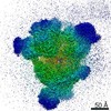 4665C 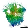 4673C  4674C 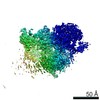 4675C 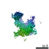 4676C 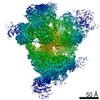 4686C  4687C 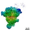 4688C 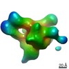 4689C  4690C 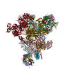 6qw6C 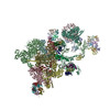 6qx9C C: 同じ文献を引用 ( |
|---|---|
| 類似構造データ | |
| 電子顕微鏡画像生データ | EMPIAR-10307 (タイトル: Human pre-B spliceosome and U4/U6.U5 tri-snRNP Data size: 2.3 TB Data #1: Dataset 1 of human pre-B spliceosome; motion-corrected micrographs [micrographs - single frame] Data #2: Dataset 2 of human pre-B spliceosome; motion-corrected micrographs [micrographs - single frame] Data #3: Dataset 3 of human pre-B spliceosome; motion-corrected micrographs [micrographs - single frame] Data #4: Dataset 4 of human pre-B spliceosome; motion-corrected micrographs [micrographs - single frame] Data #5: Dataset 5 of human pre-B spliceosome; motion-corrected micrographs [micrographs - single frame] Data #6: Selected U4/U6.U5 tri-snRNP particles after Bayesian polishing [picked particles - single frame - processed] Data #7: Crude shifted preB particles [picked particles - single frame - unprocessed]) |
-リンク
| EMDBのページ | EMDB (EBI/PDBe) / EMDataResource |
|---|
-マップ
| ファイル | ダウンロード / ファイル: emd_4672.map.gz / 形式: CCP4 / 大きさ: 282.6 MB / タイプ: IMAGE STORED AS FLOATING POINT NUMBER (4 BYTES) | ||||||||||||||||||||||||||||||||||||||||||||||||||||||||||||
|---|---|---|---|---|---|---|---|---|---|---|---|---|---|---|---|---|---|---|---|---|---|---|---|---|---|---|---|---|---|---|---|---|---|---|---|---|---|---|---|---|---|---|---|---|---|---|---|---|---|---|---|---|---|---|---|---|---|---|---|---|---|
| 注釈 | Human U5.U4/U6 tri-snRNP, multi-body refinement, Brr2 map | ||||||||||||||||||||||||||||||||||||||||||||||||||||||||||||
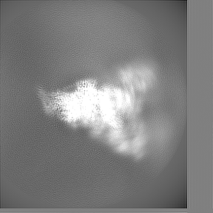
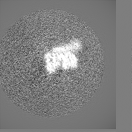
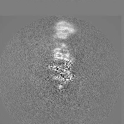
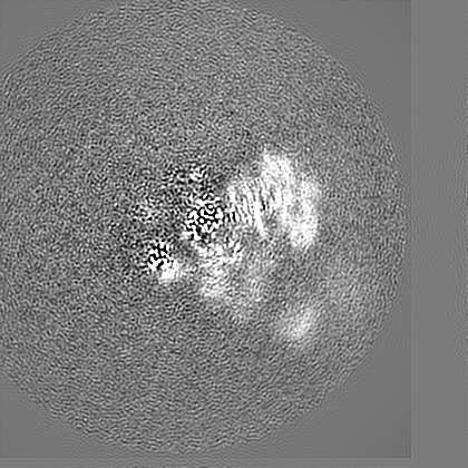
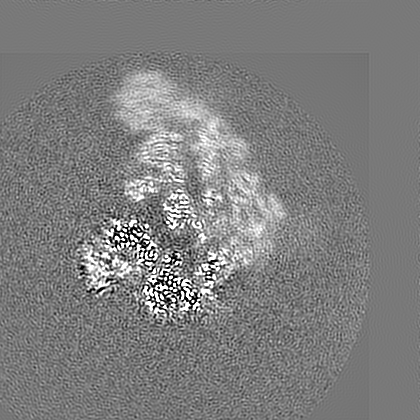
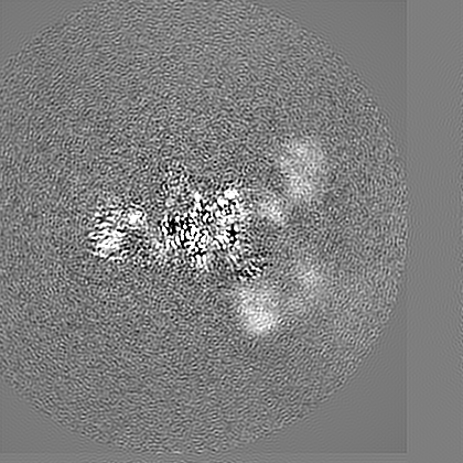
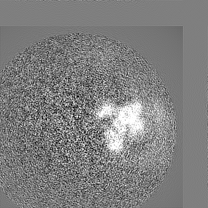
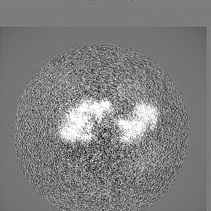
| 投影像・断面図 | 画像のコントロール
画像は Spider により作成 | ||||||||||||||||||||||||||||||||||||||||||||||||||||||||||||
| ボクセルのサイズ | X=Y=Z: 1.022 Å | ||||||||||||||||||||||||||||||||||||||||||||||||||||||||||||
| 密度 |
| ||||||||||||||||||||||||||||||||||||||||||||||||||||||||||||
| 対称性 | 空間群: 1 | ||||||||||||||||||||||||||||||||||||||||||||||||||||||||||||
| 詳細 | EMDB XML:
CCP4マップ ヘッダ情報:
| ||||||||||||||||||||||||||||||||||||||||||||||||||||||||||||
 Z (Sec.)
 Y (Row.)
 X (Col.)


-添付データ
- 試料の構成要素
-全体 : Human U4/U6.U5 tri-snRNP
| 全体 | 名称: Human U4/U6.U5 tri-snRNP |
|---|---|
| 要素 |
|
-超分子 #1: Human U4/U6.U5 tri-snRNP
| 超分子 | 名称: Human U4/U6.U5 tri-snRNP / タイプ: complex / ID: 1 / 親要素: 0 / 含まれる分子: #1-#32 |
|---|---|
| 由来(天然) | 生物種: Homo sapiens (ヒト) |
| 分子量 | 理論値: 1.7 MDa |
-実験情報
-構造解析
| 手法 | クライオ電子顕微鏡法 |
|---|---|
 解析 解析 | 単粒子再構成法 |
| 試料の集合状態 | particle |
-試料調製
| 濃度 | 1.3 mg/mL | ||||||||
|---|---|---|---|---|---|---|---|---|---|
| 緩衝液 | pH: 7.9 構成要素:
| ||||||||
| 凍結 | 凍結剤: ETHANE / チャンバー内湿度: 100 % / チャンバー内温度: 277 K / 装置: FEI VITROBOT MARK III / 詳細: Wait 30s, blot for 2s to 3s.. |
- 電子顕微鏡法
| 顕微鏡 | FEI TITAN KRIOS |
|---|---|
| 特殊光学系 | エネルギーフィルター - 名称: GIF Bioquantum / エネルギーフィルター - スリット幅: 20 eV |
| 撮影 | フィルム・検出器のモデル: GATAN K2 SUMMIT (4k x 4k) 検出モード: COUNTING / デジタル化 - 画像ごとのフレーム数: 1-40 / 平均露光時間: 6.0 sec. / 平均電子線量: 50.0 e/Å2 |
| 電子線 | 加速電圧: 300 kV / 電子線源:  FIELD EMISSION GUN FIELD EMISSION GUN |
| 電子光学系 | C2レンズ絞り径: 70.0 µm / 照射モード: FLOOD BEAM / 撮影モード: BRIGHT FIELD / Cs: 2.7 mm / 倍率(公称値): 130000 |
| 試料ステージ | 試料ホルダーモデル: FEI TITAN KRIOS AUTOGRID HOLDER ホルダー冷却材: NITROGEN |
| 実験機器 |  モデル: Titan Krios / 画像提供: FEI Company |