Movie
Movie Controller

[English] 日本語
 Yorodumi
- EMDB-45992: Structure of PDE6C in complex with the rod inhibitory p gamma sub... -
+ Open data

- Basic information
| Entry |  | |||||||||
|---|---|---|---|---|---|---|---|---|---|---|
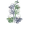
| Title | Structure of PDE6C in complex with the rod inhibitory p gamma subunit in the absence of added cGMP | |||||||||
 Map data Map data | ||||||||||
 Sample Sample |
| |||||||||
 Keywords Keywords | PDE6-cGMP-GMP complex / HYDROLASE | |||||||||
| Function / homology |  Function and homology information Function and homology informationretinal cone cell development / 3',5'-cyclic-GMP phosphodiesterase / cGMP binding / 3',5'-cyclic-GMP phosphodiesterase activity / 3',5'-cyclic-AMP phosphodiesterase activity / phototransduction, visible light / negative regulation of cAMP/PKA signal transduction / visual perception / photoreceptor disc membrane / metal ion binding / plasma membrane Similarity search - Function | |||||||||
| Biological species |  Homo sapiens (human) / Homo sapiens (human) /  | |||||||||
| Method | single particle reconstruction / cryo EM / Resolution: 3.0 Å | |||||||||
 Authors Authors | Srivastava D / Singh S / Artemyev N | |||||||||
| Funding support |  United States, 1 items United States, 1 items
| |||||||||
 Citation Citation | Journal: Proc Natl Acad Sci U S A / Year: 2025 Title: Structural and functional dynamics of human cone cGMP-phosphodiesterase important for photopic vision. Authors: Sneha Singh / Dhiraj Srivastava / Kimberly Boyd / Nikolai O Artemyev / Abstract: Cone cGMP-phosphodiesterase (PDE6) is the key effector enzyme for daylight vision, and its properties are critical for shaping distinct physiology of cone photoreceptors. We determined the structures ...Cone cGMP-phosphodiesterase (PDE6) is the key effector enzyme for daylight vision, and its properties are critical for shaping distinct physiology of cone photoreceptors. We determined the structures of human cone PDE6C in various liganded states by single-particle cryo-EM that reveal essential functional dynamics and adaptations of the enzyme. Our analysis exposed the dynamic nature of PDE6C association with its regulatory γ-subunit (Pγ) which allows openings of the catalytic pocket in the absence of phototransduction signaling, thereby controlling photoreceptor noise and sensitivity. We demonstrate evolutionarily recent adaptations of PDE6C stemming from residue substitutions in the Pγ subunit and the noncatalytic cGMP binding site and influencing the Pγ dynamics in holoPDE6C. Thus, our structural analysis sheds light on the previously unrecognized molecular evolution of the effector enzyme in cones that advances adaptation for photopic vision. | |||||||||
| History |
|
- Structure visualization
| Supplemental images |
|---|

- Downloads & links
-EMDB archive
| Map data | emd_45992.map.gz | 122.4 MB | EMDB map data format | |
|---|---|---|---|---|
| Header (meta data) | emd-45992-v30.xmlemd-45992.xml | 19.1 KB 19.1 KB | Display Display | EMDB header |
| FSC (resolution estimation) | emd_45992_fsc.xml | 10.7 KB | Display | FSC data file |
| Images |  emd_45992.png emd_45992.png | 40.7 KB | ||
| Masks | emd_45992_msk_1.map | 129.7 MB | Mask map | |
| Filedesc metadata | emd-45992.cif.gz | 6.8 KB | ||
| Others | emd_45992_half_map_1.map.gzemd_45992_half_map_2.map.gz | 120.5 MB 120.5 MB | ||
| Archive directory |  http://ftp.pdbj.org/pub/emdb/structures/EMD-45992ftp://ftp.pdbj.org/pub/emdb/structures/EMD-45992 http://ftp.pdbj.org/pub/emdb/structures/EMD-45992ftp://ftp.pdbj.org/pub/emdb/structures/EMD-45992 | HTTPS FTP |
-Related structure data
| Related structure data |  9cxiMC  9cxgC  9cxhC  9cxjC M: atomic model generated by this map C: citing same article ( |
|---|---|
| Similar structure data |


-Links
| EMDB pages | EMDB (EBI/PDBe) / EMDataResource |
|---|---|
| Related items in Molecule of the Month |


-Map
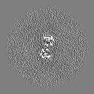
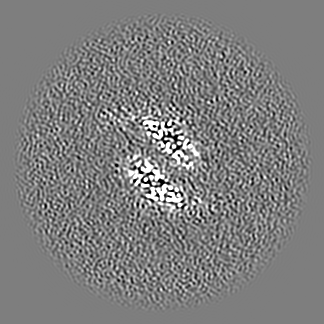
| File | Download / File: emd_45992.map.gz / Format: CCP4 / Size: 129.7 MB / Type: IMAGE STORED AS FLOATING POINT NUMBER (4 BYTES) | ||||||||||||||||||||||||||||||||||||
|---|---|---|---|---|---|---|---|---|---|---|---|---|---|---|---|---|---|---|---|---|---|---|---|---|---|---|---|---|---|---|---|---|---|---|---|---|---|
| Projections & slices | Image control
Images are generated by Spider. | ||||||||||||||||||||||||||||||||||||
| Voxel size | X=Y=Z: 0.827 Å | ||||||||||||||||||||||||||||||||||||
| Density |
| ||||||||||||||||||||||||||||||||||||
| Symmetry | Space group: 1 | ||||||||||||||||||||||||||||||||||||
| Details | EMDB XML:
|
 Z (Sec.)
 Y (Row.)
 X (Col.)


-Supplemental data
-Mask #1
| File | emd_45992_msk_1.map | ||||||||||||
|---|---|---|---|---|---|---|---|---|---|---|---|---|---|
| Projections & Slices |
| ||||||||||||
| Density Histograms |


-Half map: #2
| File | emd_45992_half_map_1.map | ||||||||||||
|---|---|---|---|---|---|---|---|---|---|---|---|---|---|
| Projections & Slices |
| ||||||||||||
| Density Histograms |


-Half map: #1
| File | emd_45992_half_map_2.map | ||||||||||||
|---|---|---|---|---|---|---|---|---|---|---|---|---|---|
| Projections & Slices |
| ||||||||||||
| Density Histograms |


- Sample components
-Entire : cone PDE6
| Entire | Name: cone PDE6 |
|---|---|
| Components |
|
-Supramolecule #1: cone PDE6
| Supramolecule | Name: cone PDE6 / type: complex / ID: 1 / Parent: 0 / Macromolecule list: #1 |
|---|---|
| Source (natural) | Organism: Homo sapiens (human) |
| Molecular weight | Theoretical: 218 KDa |
-Macromolecule #1: Cone cGMP-specific 3',5'-cyclic phosphodiesterase subunit alpha'
| Macromolecule | Name: Cone cGMP-specific 3',5'-cyclic phosphodiesterase subunit alpha' type: protein_or_peptide / ID: 1 / Number of copies: 2 / Enantiomer: LEVO / EC number: 3',5'-cyclic-GMP phosphodiesterase |
|---|---|
| Source (natural) | Organism: Homo sapiens (human) |
| Molecular weight | Theoretical: 97.724594 KDa |
| Recombinant expression | Organism:  Spodoptera aff. frugiperda 1 BOLD-2017 (butterflies/moths) Spodoptera aff. frugiperda 1 BOLD-2017 (butterflies/moths) |
| Sequence | String: GPTSGDYKDD DDKGGEINQV AVEKYLEENP QFAKEYFDRK LRVEVLGEIF KNSQVPVQSS MSFSELTQVE ESALCLELLW TVQEEGGTP EQGVHRALQR LAHLLQADRC SMFLCRSRNG IPEVASRLLD VTPTSKFEDN LVGPDKEVVF PLDIGIVGWA A HTKKTHNV ...String: GPTSGDYKDD DDKGGEINQV AVEKYLEENP QFAKEYFDRK LRVEVLGEIF KNSQVPVQSS MSFSELTQVE ESALCLELLW TVQEEGGTP EQGVHRALQR LAHLLQADRC SMFLCRSRNG IPEVASRLLD VTPTSKFEDN LVGPDKEVVF PLDIGIVGWA A HTKKTHNV PDVKKNSHFS DFMDKQTGYV TKNLLATPIV VGKEVLAVIM AVNKVNASEF SKQDEEVFSK YLNFVSIILR LH HTSYMYN IESRRSQILM WSANKVFEEL TDVERQFHKA LYTVRSYLNC ERYSIGLLDM TKEKEFYDEW PIKLGEVEPY KGP KTPDGR EVNFYKIIDY ILHGKEEIKV IPTPPADHWT LISGLPTYVA ENGFICNMMN APADEYFTFQ KGPVDETGWV IKNV LSLPI VNKKEDIVGV ATFYNRKDGK PFDEHDEYIT ETLTQFLGWS LLNTDTYDKM NKLENRKDIA QEMLMNQTKA TPEEI KSIL KFQEKLNVDV IDDCEEKQLV AILKEDLPDP RSAELYEFRF SDFPLTEHGL IKCGIRLFFE INVVEKFKVP VEVLTR WMY TVRKGYRAVT YHNWRHGFNV GQTMFTLLMT GRLKKYYTDL EAFAMLAAAF CHDIDHRGTN NLYQMKSTSP LARLHGS SI LERHHLEYSK TLLQDESLNI FQNLNKRQFE TVIHLFEVAI IATDLALYFK KRTMFQKIVD ACEQMQTEEE AIKYVTVD P TKKEIIMAMM MTACDLSAIT KPWEVQSQVA LMVANEFWEQ GDLERTVLQQ QPIPMMDRNK RDELPKLQVG FIDFVCTFV YKEFSRFHKE ITPMLSGLQN NRVEWKSLAD EYDAKMKVIE EEA UniProtKB: Cone cGMP-specific 3',5'-cyclic phosphodiesterase subunit alpha' |
-Macromolecule #2: rod pg
| Macromolecule | Name: rod pg / type: protein_or_peptide / ID: 2 / Number of copies: 2 / Enantiomer: LEVO |
|---|---|
| Source (natural) | Organism: |
| Molecular weight | Theoretical: 11.055742 KDa |
| Recombinant expression | Organism: Spodoptera aff. frugiperda 1 BOLD-2017 (butterflies/moths) |
| Sequence | String: MVGYPYDVPD YAMNLEPPKA EIRSATRVMG GPVTPRKGPP KFKQRQTRQF KSKPPKKGVQ GFGDDIPGME GLGTDITVIC PWEAFNHLE LHELAQYGII |
-Macromolecule #3: ZINC ION
| Macromolecule | Name: ZINC ION / type: ligand / ID: 3 / Number of copies: 2 / Formula: ZN |
|---|---|
| Molecular weight | Theoretical: 65.409 Da |
-Macromolecule #4: MAGNESIUM ION
| Macromolecule | Name: MAGNESIUM ION / type: ligand / ID: 4 / Number of copies: 2 / Formula: MG |
|---|---|
| Molecular weight | Theoretical: 24.305 Da |
-Macromolecule #5: CYCLIC GUANOSINE MONOPHOSPHATE
| Macromolecule | Name: CYCLIC GUANOSINE MONOPHOSPHATE / type: ligand / ID: 5 / Number of copies: 2 / Formula: PCG |
|---|---|
| Molecular weight | Theoretical: 345.205 Da |
| Chemical component information |  ChemComp-PCG: |
-Experimental details
-Structure determination
| Method | cryo EM |
|---|---|
 Processing Processing | single particle reconstruction |
| Aggregation state | particle |
-Sample preparation
| Concentration | 0.5 mg/mL |
|---|---|
| Buffer | pH: 7.5 |
| Vitrification | Cryogen name: ETHANE |
- Electron microscopy
| Microscope | FEI TITAN KRIOS |
|---|---|
| Image recording | Film or detector model: GATAN K3 (6k x 4k) / Average electron dose: 60.0 e/Å2 |
| Electron beam | Acceleration voltage: 300 kV / Electron source:  FIELD EMISSION GUN FIELD EMISSION GUN |
| Electron optics | Illumination mode: FLOOD BEAM / Imaging mode: OTHER / Nominal defocus max: 3.0 µm / Nominal defocus min: 1.0 µm / Nominal magnification: 105000 |
| Sample stage | Specimen holder model: FEI TITAN KRIOS AUTOGRID HOLDER / Cooling holder cryogen: NITROGEN |
| Experimental equipment |  Model: Titan Krios / Image courtesy: FEI Company |