National Institutes of Health/National Institute of Dental and Craniofacial Research (NIH/NIDCR)
R01DE025567
米国
引用
ジャーナル: J Virol / 年: 2025 タイトル: Structure of the Kaposi's sarcoma-associated herpesvirus gB in post-fusion conformation. 著者: Fumiaki Ito / James Zhen / Guodong Xie / Haigen Huang / Juan C Silva / Ting-Ting Wu / Z Hong Zhou / 要旨: Discovered in 1994 in lesions of an AIDS patient, Kaposi's sarcoma-associated herpesvirus (KSHV) is a member of the gammaherpesvirus subfamily of the family, which contains a total of nine that ...Discovered in 1994 in lesions of an AIDS patient, Kaposi's sarcoma-associated herpesvirus (KSHV) is a member of the gammaherpesvirus subfamily of the family, which contains a total of nine that infect humans. These viruses all contain a large envelope glycoprotein, glycoprotein B (gB), that is required for viral fusion with host cell membrane to initial infection. Although the atomic structures of five other human herpesviruses in their postfusion conformation and one in its prefusion conformation are known, the atomic structure of KSHV gB has not been reported. Here, we report the first structure of the KSHV gB ectodomain determined by single-particle cryogenic electron microscopy (cryoEM). Despite a similar global fold between herpesvirus gB, KSHV gB possesses local differences not shared by its relatives in other herpesviruses. The glycosylation sites of gB are arranged in belts down the symmetry axis with distinct localization compared to that of other herpesviruses, which occludes certain antibody binding sites. An extended glycan chain observed in domain I (DI), located proximal to the host membrane, may suggest its possible role in host cell attachment. Local flexibility of domain IV (DIV) governed by molecular hinges at its interdomain junctions identifies a means for enabling conformational change. A mutation in the domain III (DIII) central helix disrupts incorporation of gB into KSHV virions despite adoption of a canonical fold . Taken together, this study reveals mechanisms of structural variability of herpesvirus fusion protein gB and informs its folding and immunogenicity.IMPORTANCEIn 1994, a cancer-causing virus was discovered in lesions of AIDS patients, which was later named Kaposi's sarcoma-associated herpesvirus (KSHV). As the latest discovered human herpesvirus, KSHV has been classified into the gammaherpesvirus subfamily of the . In this study, we have expressed KSHV gB and employed cryogenic electron microscopy (cryoEM) to determine its first structure. Importantly, our structure resolves some glycans beyond the first sugar moiety. These glycans are arranged in a pattern unique to KSHV, which impacts the antigenicity of KSHV gB. Our structure also reveals conformational flexibility caused by molecular hinges between domains that provide clues into the mechanism behind the drastic change between prefusion and postfusion states.
 ムービー
ムービー コントローラー


 データを開く
 基本情報
基本情報
 マップデータ
 試料
試料 キーワード
キーワード 機能・相同性情報
 Human gammaherpesvirus 8 (ヘルペスウイルス)
 データ登録者
 米国, 1件
  引用
引用 構造の表示
構造の表示
 ダウンロードとリンク
 emd_45927.png
 http://ftp.pdbj.org/pub/emdb/structures/EMD-45927
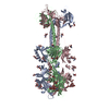
 Z (Sec.)
 Y (Row.)
 X (Col.)


 試料の構成要素
 Cricetulus griseus (モンゴルキヌゲネズミ)
 解析
解析 電子顕微鏡法
 FIELD EMISSION GUN

