National Institutes of Health/National Institute of Dental and Craniofacial Research (NIH/NIDCR)
R01DE025567
United States
Citation
Journal: J Virol / Year: 2025 Title: Structure of the Kaposi's sarcoma-associated herpesvirus gB in post-fusion conformation. Authors: Fumiaki Ito / James Zhen / Guodong Xie / Haigen Huang / Juan C Silva / Ting-Ting Wu / Z Hong Zhou / Abstract: Discovered in 1994 in lesions of an AIDS patient, Kaposi's sarcoma-associated herpesvirus (KSHV) is a member of the gammaherpesvirus subfamily of the family, which contains a total of nine that ...Discovered in 1994 in lesions of an AIDS patient, Kaposi's sarcoma-associated herpesvirus (KSHV) is a member of the gammaherpesvirus subfamily of the family, which contains a total of nine that infect humans. These viruses all contain a large envelope glycoprotein, glycoprotein B (gB), that is required for viral fusion with host cell membrane to initial infection. Although the atomic structures of five other human herpesviruses in their postfusion conformation and one in its prefusion conformation are known, the atomic structure of KSHV gB has not been reported. Here, we report the first structure of the KSHV gB ectodomain determined by single-particle cryogenic electron microscopy (cryoEM). Despite a similar global fold between herpesvirus gB, KSHV gB possesses local differences not shared by its relatives in other herpesviruses. The glycosylation sites of gB are arranged in belts down the symmetry axis with distinct localization compared to that of other herpesviruses, which occludes certain antibody binding sites. An extended glycan chain observed in domain I (DI), located proximal to the host membrane, may suggest its possible role in host cell attachment. Local flexibility of domain IV (DIV) governed by molecular hinges at its interdomain junctions identifies a means for enabling conformational change. A mutation in the domain III (DIII) central helix disrupts incorporation of gB into KSHV virions despite adoption of a canonical fold . Taken together, this study reveals mechanisms of structural variability of herpesvirus fusion protein gB and informs its folding and immunogenicity.IMPORTANCEIn 1994, a cancer-causing virus was discovered in lesions of AIDS patients, which was later named Kaposi's sarcoma-associated herpesvirus (KSHV). As the latest discovered human herpesvirus, KSHV has been classified into the gammaherpesvirus subfamily of the . In this study, we have expressed KSHV gB and employed cryogenic electron microscopy (cryoEM) to determine its first structure. Importantly, our structure resolves some glycans beyond the first sugar moiety. These glycans are arranged in a pattern unique to KSHV, which impacts the antigenicity of KSHV gB. Our structure also reveals conformational flexibility caused by molecular hinges between domains that provide clues into the mechanism behind the drastic change between prefusion and postfusion states.
In the structure databanks used in Yorodumi, some data are registered as the other names, "COVID-19 virus" and "2019-nCoV". Here are the details of the virus and the list of structure data.
Jan 31, 2019. EMDB accession codes are about to change! (news from PDBe EMDB page)
EMDB accession codes are about to change! (news from PDBe EMDB page)
The allocation of 4 digits for EMDB accession codes will soon come to an end. Whilst these codes will remain in use, new EMDB accession codes will include an additional digit and will expand incrementally as the available range of codes is exhausted. The current 4-digit format prefixed with “EMD-” (i.e. EMD-XXXX) will advance to a 5-digit format (i.e. EMD-XXXXX), and so on. It is currently estimated that the 4-digit codes will be depleted around Spring 2019, at which point the 5-digit format will come into force.
The EM Navigator/Yorodumi systems omit the EMD- prefix.
Related info.:Q: What is EMD? / ID/Accession-code notation in Yorodumi/EM Navigator
Yorodumi is a browser for structure data from EMDB, PDB, SASBDB, etc.
This page is also the successor to EM Navigator detail page, and also detail information page/front-end page for Omokage search.
The word "yorodu" (or yorozu) is an old Japanese word meaning "ten thousand". "mi" (miru) is to see.
Related info.:EMDB / PDB / SASBDB / Comparison of 3 databanks / Yorodumi Search / Aug 31, 2016. New EM Navigator & Yorodumi / Yorodumi Papers / Jmol/JSmol / Function and homology information / Changes in new EM Navigator and Yorodumi
 Movie
Movie Controller


 Open data
 Basic information
 Map data
 Sample
 Keywords
 Function and homology information
 Human gammaherpesvirus 8
 Authors
 United States, 1 items
  Citation
 Structure visualization
 Downloads & links
 emd_45927.png
 http://ftp.pdbj.org/pub/emdb/structures/EMD-45927
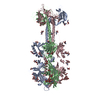
 Z (Sec.)
 Y (Row.)
 X (Col.)


 Sample components
 Cricetulus griseus (Chinese hamster)
 Processing
 Electron microscopy
 FIELD EMISSION GUN

