 Movie
Movie Controller

[English] 日本語
 Yorodumi
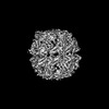
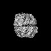
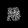
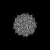
- EMDB-45886: TRiC-ADP-S2 state is a conformation when TRiC incubated in 1 mM ADP -
+ Open data

- Basic information
| Entry | 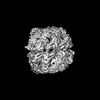 | |||||||||
|---|---|---|---|---|---|---|---|---|---|---|
| Title | TRiC-ADP-S2 state is a conformation when TRiC incubated in 1 mM ADP | |||||||||
 Map data Map data | TRiC-ADP-S2 | |||||||||
 Sample Sample |
| |||||||||
 Keywords Keywords | Complex / Chaperonin / ADP / CHAPERONE | |||||||||
| Function / homology |  Function and homology information Function and homology informationAssociation of TriC/CCT with target proteins during biosynthesis / Cooperation of PDCL (PhLP1) and TRiC/CCT in G-protein beta folding / chaperonin-containing T-complex / Neutrophil degranulation / ATP-dependent protein folding chaperone / : / protein folding / ATP hydrolysis activity / ATP binding / plasma membrane / cytoplasm Similarity search - Function | |||||||||
| Biological species |  | |||||||||
| Method | single particle reconstruction / cryo EM / Resolution: 5.6 Å | |||||||||
 Authors Authors | Jin M / Cong Y | |||||||||
| Funding support |  China, 2 items China, 2 items
| |||||||||
 Citation Citation | Journal: QRB Discov / Year: 2025 Title: The conformational landscape of TRiC ring-opening and its underlying stepwise mechanism revealed by cryo-EM. Authors: Mingliang Jin / Yunxiang Zang / Huping Wang / Yao Cong / Abstract: The TRiC/CCT complex assists in the folding of approximately 10% of cytosolic proteins through an ATP-driven conformational cycle, playing a crucial role in maintaining protein homeostasis. Despite ...The TRiC/CCT complex assists in the folding of approximately 10% of cytosolic proteins through an ATP-driven conformational cycle, playing a crucial role in maintaining protein homeostasis. Despite our understanding of ATP-driven TRiC ring closing and substrate folding, the process and mechanisms underlying TRiC ring-opening and substrate release remain largely unexplored. In this study, by determining an ensemble of cryo-EM structures of yeast TRiC in the presence of ADP, including three intermediate transition states, we present a comprehensive picture of the TRiC ring-opening process. During this process, CCT3 detects the loss of γ-phosphate and initiates with the dynamics of its apical protrusion, and expands to the outward leaning of the consecutive CCT6/8/7/5 subunits. This is followed by significant movements of CCT2, CCT4, and especially CCT1 subunits, resulting in the opening of the TRiC rings. We also observed an unforeseen temporary separation between the two rings in the CCT2 side, coordinating the release of the originally locked CCT4 N-terminus, which potentially participates in the ring-opening process. Collectively, our study reveals a stepwise TRiC ring-opening mechanism, provides a comprehensive view of the TRiC conformational landscape, and sheds lights on its subunit specificity in sensing nucleotide status and substrate release. Our findings deepen our understanding of protein folding assisted by TRiC and may inspire new strategies for the diagnosis and treatment of related diseases. | |||||||||
| History |
|
- Structure visualization
| Supplemental images |
|---|

- Downloads & links
-EMDB archive
| Map data | emd_45886.map.gz | 4 MB | EMDB map data format | |
|---|---|---|---|---|
| Header (meta data) | emd-45886-v30.xmlemd-45886.xml | 23.9 KB 23.9 KB | Display Display | EMDB header |
| Images |  emd_45886.png emd_45886.png | 106.1 KB | ||
| Filedesc metadata | emd-45886.cif.gz | 9.1 KB | ||
| Archive directory |  http://ftp.pdbj.org/pub/emdb/structures/EMD-45886ftp://ftp.pdbj.org/pub/emdb/structures/EMD-45886 http://ftp.pdbj.org/pub/emdb/structures/EMD-45886ftp://ftp.pdbj.org/pub/emdb/structures/EMD-45886 | HTTPS FTP |
-Related structure data
| Related structure data | 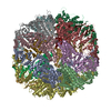 9cs3MC  9cr2C  9cs4C 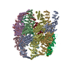 9cs6C 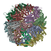 9csaC M: atomic model generated by this map C: citing same article ( |
|---|---|
| Similar structure data |

-Links
| EMDB pages | EMDB (EBI/PDBe) / EMDataResource |
|---|---|
| Related items in Molecule of the Month |


-Map
| File | Download / File: emd_45886.map.gz / Format: CCP4 / Size: 64 MB / Type: IMAGE STORED AS FLOATING POINT NUMBER (4 BYTES) | ||||||||||||||||||||||||||||||||||||
|---|---|---|---|---|---|---|---|---|---|---|---|---|---|---|---|---|---|---|---|---|---|---|---|---|---|---|---|---|---|---|---|---|---|---|---|---|---|
| Annotation | TRiC-ADP-S2 | ||||||||||||||||||||||||||||||||||||
| Projections & slices | Image control
Images are generated by Spider. | ||||||||||||||||||||||||||||||||||||
| Voxel size | X=Y=Z: 1.318 Å | ||||||||||||||||||||||||||||||||||||
| Density |
| ||||||||||||||||||||||||||||||||||||
| Symmetry | Space group: 1 | ||||||||||||||||||||||||||||||||||||
| Details | EMDB XML:
|
 Z (Sec.)
 Y (Row.)
 X (Col.)


-Supplemental data
- Sample components
+Entire : TRiC at ADP state 2
+Supramolecule #1: TRiC at ADP state 2
+Supramolecule #2: T-complex protein 1 subunit gamma
+Supramolecule #3: the other TRiC subunits
+Macromolecule #1: T-complex protein 1 subunit alpha
+Macromolecule #2: T-complex protein 1 subunit beta
+Macromolecule #3: T-complex protein 1 subunit epsilon
+Macromolecule #4: T-complex protein 1 subunit gamma
+Macromolecule #5: T-complex protein 1 subunit eta
+Macromolecule #6: T-complex protein 1 subunit theta
+Macromolecule #7: T-complex protein 1 subunit zeta
+Macromolecule #8: T-complex protein 1 subunit delta
-Experimental details
-Structure determination
| Method | cryo EM |
|---|---|
 Processing Processing | single particle reconstruction |
| Aggregation state | particle |
-Sample preparation
| Buffer | pH: 7.4 |
|---|---|
| Vitrification | Cryogen name: ETHANE |
- Electron microscopy
| Microscope | TFS KRIOS |
|---|---|
| Image recording | Film or detector model: GATAN K2 SUMMIT (4k x 4k) / Average electron dose: 38.0 e/Å2 |
| Electron beam | Acceleration voltage: 300 kV / Electron source:  FIELD EMISSION GUN FIELD EMISSION GUN |
| Electron optics | Illumination mode: FLOOD BEAM / Imaging mode: BRIGHT FIELD / Nominal defocus max: 2.4 µm / Nominal defocus min: 1.2 µm |
| Experimental equipment |  Model: Titan Krios / Image courtesy: FEI Company |
