 Movie
Movie Controller

[English] 日本語
 Yorodumi
- EMDB-45671: Subtomogram average of the Polar Tube Outer Filament layer from E... -
+ Open data

- Basic information
| Entry | 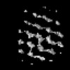 | |||||||||||||||||||||||||||
|---|---|---|---|---|---|---|---|---|---|---|---|---|---|---|---|---|---|---|---|---|---|---|---|---|---|---|---|---|
| Title | Subtomogram average of the Polar Tube Outer Filament layer from Encephalitozoon intestinalis microsporidian spores | |||||||||||||||||||||||||||
 Map data Map data | Filtered map | |||||||||||||||||||||||||||
 Sample Sample |
| |||||||||||||||||||||||||||
 Keywords Keywords | Microsporidia / polar tube / subtomogram averaging / cryo-ET / CELL INVASION | |||||||||||||||||||||||||||
| Biological species |  Encephalitozoon intestinalis (fungus) Encephalitozoon intestinalis (fungus) | |||||||||||||||||||||||||||
| Method | subtomogram averaging / cryo EM / Resolution: 36.0 Å | |||||||||||||||||||||||||||
 Authors Authors | Usmani M / Coudray N / Bobe D / Kopylov M / Ekiert DC / Bhabha G | |||||||||||||||||||||||||||
| Funding support |  United States, 8 items United States, 8 items
| |||||||||||||||||||||||||||
 Citation Citation | Journal: bioRxiv / Year: 2024 Title: Cryo-ET reveals the architecture of the polar tube invasion apparatus from microsporidian parasites. Authors: Mahrukh Usmani / Nicolas Coudray / Margot Riggi / Rishwanth Raghu / Harshita Ramchandani / Daija Bobe / Mykhailo Kopylov / Ellen D Zhong / Janet H Iwasa / Damian C Ekiert / Gira Bhabha Abstract: Microsporidia are divergent fungal pathogens that employ a harpoon-like apparatus called the polar tube (PT) to invade host cells. The PT architecture and its association with neighboring organelles ...Microsporidia are divergent fungal pathogens that employ a harpoon-like apparatus called the polar tube (PT) to invade host cells. The PT architecture and its association with neighboring organelles remain poorly understood. Here, we use cryo-electron tomography to investigate the structural cell biology of the PT in dormant spores from the human-infecting microsporidian species, . Segmentation and subtomogram averaging of the PT reveal at least four layers: two protein-based layers surrounded by a membrane, and filled with a dense core. Regularly spaced protein filaments form the structural skeleton of the PT. Combining cryo-electron tomography with cellular modeling, we propose a model for the 3-dimensional organization of the polaroplast, an organelle that is continuous with the membrane layer that envelops the PT. Our results reveal the ultrastructure of the microsporidian invasion apparatus , laying the foundation for understanding infection mechanisms. | |||||||||||||||||||||||||||
| History |
|
- Structure visualization
| Supplemental images |
|---|

- Downloads & links
-EMDB archive
| Map data | emd_45671.map.gz | 481.6 KB |  EMDB map data format EMDB map data format | |
|---|---|---|---|---|
| Header (meta data) | emd-45671-v30.xmlemd-45671.xml | 22.3 KB 22.3 KB | Display Display | EMDB header |
| Images |  emd_45671.png emd_45671.png | 39.9 KB | ||
| Masks | emd_45671_msk_1.map | 1 MB | Mask map | |
| Filedesc metadata | emd-45671.cif.gz | 5.4 KB | ||
| Others | emd_45671_additional_1.map.gzemd_45671_half_map_1.map.gzemd_45671_half_map_2.map.gz | 730.1 KB 736 KB 735.8 KB | ||
| Archive directory |  http://ftp.pdbj.org/pub/emdb/structures/EMD-45671ftp://ftp.pdbj.org/pub/emdb/structures/EMD-45671 http://ftp.pdbj.org/pub/emdb/structures/EMD-45671ftp://ftp.pdbj.org/pub/emdb/structures/EMD-45671 | HTTPS FTP |
-Related structure data
-Links
| EMDB pages | EMDB (EBI/PDBe) / EMDataResource |
|---|
-Map
| File | Download / File: emd_45671.map.gz / Format: CCP4 / Size: 1 MB / Type: IMAGE STORED AS FLOATING POINT NUMBER (4 BYTES) | ||||||||||||||||||||||||||||||||||||
|---|---|---|---|---|---|---|---|---|---|---|---|---|---|---|---|---|---|---|---|---|---|---|---|---|---|---|---|---|---|---|---|---|---|---|---|---|---|
| Annotation | Filtered map | ||||||||||||||||||||||||||||||||||||
| Projections & slices | Image control
Images are generated by Spider. | ||||||||||||||||||||||||||||||||||||
| Voxel size | X=Y=Z: 6.862 Å | ||||||||||||||||||||||||||||||||||||
| Density |
| ||||||||||||||||||||||||||||||||||||
| Symmetry | Space group: 1 | ||||||||||||||||||||||||||||||||||||
| Details | EMDB XML:
|
 Z (Sec.)
 Y (Row.)
 X (Col.)


-Supplemental data
-Mask #1
| File | emd_45671_msk_1.map | ||||||||||||
|---|---|---|---|---|---|---|---|---|---|---|---|---|---|
| Projections & Slices |
| ||||||||||||
| Density Histograms |


-Additional map: Raw unfiltered map
| File | emd_45671_additional_1.map | ||||||||||||
|---|---|---|---|---|---|---|---|---|---|---|---|---|---|
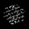
| Annotation | Raw unfiltered map | ||||||||||||
| Projections & Slices |
| ||||||||||||
| Density Histograms |


-Half map: #2
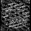
| File | emd_45671_half_map_1.map | ||||||||||||
|---|---|---|---|---|---|---|---|---|---|---|---|---|---|
| Projections & Slices |
| ||||||||||||
| Density Histograms |


-Half map: #1
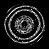
| File | emd_45671_half_map_2.map | ||||||||||||
|---|---|---|---|---|---|---|---|---|---|---|---|---|---|
| Projections & Slices |
| ||||||||||||
| Density Histograms |


- Sample components
-Entire : Encephalitozoon intestinalis microsporidian spores
| Entire | Name: Encephalitozoon intestinalis microsporidian spores |
|---|---|
| Components |
|
-Supramolecule #1: Encephalitozoon intestinalis microsporidian spores
| Supramolecule | Name: Encephalitozoon intestinalis microsporidian spores / type: cell / ID: 1 / Parent: 0 |
|---|---|
| Source (natural) | Organism: Encephalitozoon intestinalis (fungus) |
-Experimental details
-Structure determination
| Method | cryo EM |
|---|---|
 Processing Processing | subtomogram averaging |
| Aggregation state | cell |
-Sample preparation
| Buffer | pH: 7 Component:
Details: Buffer details: DPBS (Gibco 14190144) | |||||||||||||||
|---|---|---|---|---|---|---|---|---|---|---|---|---|---|---|---|---|
| Grid | Model: Quantifoil R2/2 / Material: COPPER / Mesh: 200 / Support film - Material: CARBON / Support film - topology: CONTINUOUS / Support film - Film thickness: 20 / Pretreatment - Type: GLOW DISCHARGE / Pretreatment - Time: 30 sec. | |||||||||||||||
| Vitrification | Cryogen name: NITROGEN / Details: Wohlwend Compact 01 HPF. | |||||||||||||||
| Details | 1.3e8 spores per uL |
- Electron microscopy
| Microscope | FEI TITAN KRIOS |
|---|---|
| Image recording | Film or detector model: GATAN K3 (6k x 4k) / Digitization - Dimensions - Width: 5760 pixel / Digitization - Dimensions - Height: 4092 pixel / Number real images: 1 / Average exposure time: 1.0 sec. / Average electron dose: 2.87 e/Å2 |
| Electron beam | Acceleration voltage: 300 kV / Electron source:  FIELD EMISSION GUN FIELD EMISSION GUN |
| Electron optics | Illumination mode: FLOOD BEAM / Imaging mode: BRIGHT FIELD / Cs: 2.7 mm / Nominal defocus max: 5.5 µm / Nominal defocus min: 4.0 µm / Nominal magnification: 26000 |
| Sample stage | Specimen holder model: FEI TITAN KRIOS AUTOGRID HOLDER |
| Experimental equipment |  Model: Titan Krios / Image courtesy: FEI Company |
-Image processing
| Final reconstruction | Number classes used: 1 / Applied symmetry - Point group: C1 (asymmetric) / Algorithm: BACK PROJECTION / Resolution.type: BY AUTHOR / Resolution: 36.0 Å / Resolution method: FSC 0.143 CUT-OFF / Software - Name: RELION / Number subtomograms used: 1657 |
|---|---|
| Extraction | Number tomograms: 1 / Number images used: 7353 / Software - Name: Dynamo |
| Final 3D classification | Software - Name: RELION |
| Final angle assignment | Type: MAXIMUM LIKELIHOOD / Software - Name: RELION |
