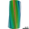
[English] 日本語
Yorodumi- EMDB-45465: Structure of type I-2 alpha-synuclein filament from multiple syst... -
+
Open data
-
Basic information
| Entry | ![]() | ||||||||||||
|---|---|---|---|---|---|---|---|---|---|---|---|---|---|
| Title | Structure of type I-2 alpha-synuclein filament from multiple system atrophy | ||||||||||||
Map data | |||||||||||||
Sample |
| ||||||||||||
Keywords | fibril / synuclein / PROTEIN FIBRIL | ||||||||||||
| Function / homology | Function and homology informationnegative regulation of mitochondrial electron transport, NADH to ubiquinone / : / neutral lipid metabolic process / regulation of acyl-CoA biosynthetic process / negative regulation of dopamine uptake involved in synaptic transmission / negative regulation of norepinephrine uptake / response to desipramine / positive regulation of SNARE complex assembly / positive regulation of hydrogen peroxide catabolic process / supramolecular fiber ...negative regulation of mitochondrial electron transport, NADH to ubiquinone / : / neutral lipid metabolic process / regulation of acyl-CoA biosynthetic process / negative regulation of dopamine uptake involved in synaptic transmission / negative regulation of norepinephrine uptake / response to desipramine / positive regulation of SNARE complex assembly / positive regulation of hydrogen peroxide catabolic process / supramolecular fiber / regulation of synaptic vesicle recycling / negative regulation of chaperone-mediated autophagy / mitochondrial membrane organization / regulation of reactive oxygen species biosynthetic process / negative regulation of platelet-derived growth factor receptor signaling pathway / positive regulation of protein localization to cell periphery / negative regulation of exocytosis / regulation of glutamate secretion / dopamine biosynthetic process / response to iron(II) ion / SNARE complex assembly / negative regulation of dopamine metabolic process / positive regulation of neurotransmitter secretion / regulation of macrophage activation / positive regulation of inositol phosphate biosynthetic process / regulation of norepinephrine uptake / negative regulation of microtubule polymerization / regulation of locomotion / synaptic vesicle transport / transporter regulator activity / synaptic vesicle priming / dopamine uptake involved in synaptic transmission / protein kinase inhibitor activity / regulation of dopamine secretion / negative regulation of thrombin-activated receptor signaling pathway / mitochondrial ATP synthesis coupled electron transport / dynein complex binding / positive regulation of receptor recycling / cuprous ion binding / nuclear outer membrane / response to magnesium ion / positive regulation of exocytosis / synaptic vesicle exocytosis / positive regulation of endocytosis / kinesin binding / synaptic vesicle endocytosis / enzyme inhibitor activity / cysteine-type endopeptidase inhibitor activity / regulation of presynapse assembly / response to type II interferon / negative regulation of serotonin uptake / alpha-tubulin binding / beta-tubulin binding / phospholipase binding / behavioral response to cocaine / supramolecular fiber organization / phospholipid metabolic process / cellular response to fibroblast growth factor stimulus / axon terminus / inclusion body / cellular response to epinephrine stimulus / Hsp70 protein binding / response to interleukin-1 / regulation of microtubule cytoskeleton organization / cellular response to copper ion / positive regulation of release of sequestered calcium ion into cytosol / SNARE binding / adult locomotory behavior / excitatory postsynaptic potential / protein tetramerization / phosphoprotein binding / microglial cell activation / ferrous iron binding / fatty acid metabolic process / regulation of long-term neuronal synaptic plasticity / synapse organization / PKR-mediated signaling / protein destabilization / phospholipid binding / receptor internalization / tau protein binding / long-term synaptic potentiation / terminal bouton / positive regulation of inflammatory response / synaptic vesicle membrane / actin cytoskeleton / growth cone / actin binding / cellular response to oxidative stress / neuron apoptotic process / cell cortex / histone binding / response to lipopolysaccharide / microtubule binding / chemical synaptic transmission / amyloid fibril formation / molecular adaptor activity / negative regulation of neuron apoptotic process / mitochondrial outer membrane / oxidoreductase activity Similarity search - Function | ||||||||||||
| Biological species | Homo sapiens (human) | ||||||||||||
| Method | helical reconstruction / cryo EM / Resolution: 3.3 Å | ||||||||||||
Authors | Yan NL / Candido F / Tse E / Melo AA / Southworth DR / Merz GE | ||||||||||||
| Funding support | United States, 3 items
| ||||||||||||
Citation | Journal: FEBS Lett / Year: 2025Title: Cryo-EM structure of a novel α-synuclein filament subtype from multiple system atrophy. Authors: Nicholas L Yan / Francisco Candido / Eric Tse / Arthur A Melo / Stanley B Prusiner / Daniel A Mordes / Daniel R Southworth / Nick A Paras / Gregory E Merz / ![]() Abstract: Multiple system atrophy (MSA) is a progressive neurodegenerative disease characterized by accumulation of α-synuclein cross-β amyloid filaments in the brain. Previous structural studies of these ...Multiple system atrophy (MSA) is a progressive neurodegenerative disease characterized by accumulation of α-synuclein cross-β amyloid filaments in the brain. Previous structural studies of these filaments by cryo-electron microscopy (cryo-EM) revealed three discrete folds distinct from α-synuclein filaments associated with other neurodegenerative diseases. Here, we use cryo-EM to identify a novel, low-populated MSA filament subtype (designated Type I) in addition to a predominant class comprising MSA Type II filaments. The 3.3-Å resolution structure of the Type I filament reveals a fold consisting of two asymmetric protofilaments, one of which adopts a novel structure that is chimeric between two previously reported protofilaments. These results further define MSA-specific folds of α-synuclein filaments and have implications for designing MSA diagnostics and therapeutics. | ||||||||||||
| History |
|
-
Structure visualization
| Supplemental images |
|---|
-
Downloads & links
-EMDB archive
| Map data | emd_45465.map.gz | 7.9 MB | EMDB map data format | |
|---|---|---|---|---|
| Header (meta data) | emd-45465-v30.xml emd-45465.xml | 17.1 KB 17.1 KB | Display Display | EMDB header |
| FSC (resolution estimation) | emd_45465_fsc.xml | 10.2 KB | Display | FSC data file |
| Images | emd_45465.png | 102.8 KB | ||
| Filedesc metadata | emd-45465.cif.gz | 5.9 KB | ||
| Others | emd_45465_half_map_1.map.gz emd_45465_half_map_2.map.gz | 71.3 MB 71.3 MB | ||
| Archive directory | http://ftp.pdbj.org/pub/emdb/structures/EMD-45465 ftp://ftp.pdbj.org/pub/emdb/structures/EMD-45465 | HTTPS FTP |
-Validation report
| Summary document | emd_45465_validation.pdf.gz | 901.7 KB | Display | EMDB validaton report |
|---|---|---|---|---|
| Full document | emd_45465_full_validation.pdf.gz | 901.3 KB | Display | |
| Data in XML | emd_45465_validation.xml.gz | 17.5 KB | Display | |
| Data in CIF | emd_45465_validation.cif.gz | 23 KB | Display | |
| Arichive directory | https://ftp.pdbj.org/pub/emdb/validation_reports/EMD-45465 ftp://ftp.pdbj.org/pub/emdb/validation_reports/EMD-45465 | HTTPS FTP |
-Related structure data
| Related structure data | ![]() 9cdaMC ![]() 9cd9C C: citing same article ( M: atomic model generated by this map |
|---|---|
| Similar structure data | Similarity search - Function & homology F&H Search |
-
Links
| EMDB pages | EMDB (EBI/PDBe) / EMDataResource |
|---|
-
Map
| File | Download / File: emd_45465.map.gz / Format: CCP4 / Size: 91.1 MB / Type: IMAGE STORED AS FLOATING POINT NUMBER (4 BYTES) | ||||||||||||||||||||||||||||||||||||
|---|---|---|---|---|---|---|---|---|---|---|---|---|---|---|---|---|---|---|---|---|---|---|---|---|---|---|---|---|---|---|---|---|---|---|---|---|---|
| Projections & slices | Image control
Images are generated by Spider. | ||||||||||||||||||||||||||||||||||||
| Voxel size | X=Y=Z: 0.834 Å | ||||||||||||||||||||||||||||||||||||
| Density |
| ||||||||||||||||||||||||||||||||||||
| Symmetry | Space group: 1 | ||||||||||||||||||||||||||||||||||||
| Details | EMDB XML:
|
-Supplemental data
-Half map: #1
| File | emd_45465_half_map_1.map | ||||||||||||
|---|---|---|---|---|---|---|---|---|---|---|---|---|---|
| Projections & Slices |
| ||||||||||||
| Density Histograms |
-Half map: #2
| File | emd_45465_half_map_2.map | ||||||||||||
|---|---|---|---|---|---|---|---|---|---|---|---|---|---|
| Projections & Slices |
| ||||||||||||
| Density Histograms |
-
Sample components
-Entire : Type I-2 alpha-synuclein filament from multiple system atrophy
| Entire | Name: Type I-2 alpha-synuclein filament from multiple system atrophy |
|---|---|
| Components |
|
-Supramolecule #1: Type I-2 alpha-synuclein filament from multiple system atrophy
| Supramolecule | Name: Type I-2 alpha-synuclein filament from multiple system atrophy type: complex / ID: 1 / Parent: 0 / Macromolecule list: all |
|---|---|
| Source (natural) | Organism: Homo sapiens (human) / Organ: Brain / Tissue: Cerebellum |
-Macromolecule #1: Alpha-synuclein
| Macromolecule | Name: Alpha-synuclein / type: protein_or_peptide / ID: 1 / Number of copies: 18 / Enantiomer: LEVO |
|---|---|
| Source (natural) | Organism: Homo sapiens (human) / Organ: Brain / Tissue: Cerebellum |
| Molecular weight | Theoretical: 14.476108 KDa |
| Sequence | String: MDVFMKGLSK AKEGVVAAAE KTKQGVAEAA GKTKEGVLYV GSKTKEGVVH GVATVAEKTK EQVTNVGGAV VTGVTAVAQK TVEGAGSIA AATGFVKKDQ LGKNEEGAPQ EGILEDMPVD PDNEAYEMPS EEGYQDYEPE A UniProtKB: Alpha-synuclein |
-Experimental details
-Structure determination
| Method | cryo EM |
|---|---|
Processing | helical reconstruction |
| Aggregation state | filament |
-
Sample preparation
| Buffer | pH: 7.4 / Component - Concentration: 30.0 mM / Component - Name: Tris-HCl |
|---|---|
| Grid | Model: Quantifoil R1.2/1.3 / Material: GOLD / Mesh: 200 / Support film - Material: CARBON / Support film - topology: HOLEY |
| Vitrification | Cryogen name: ETHANE / Chamber humidity: 100 % / Chamber temperature: 277 K / Instrument: FEI VITROBOT MARK IV / Details: Wait time 30s, blot time 7.5s. |
-
Electron microscopy
| Microscope | TFS KRIOS |
|---|---|
| Image recording | Film or detector model: GATAN K3 (6k x 4k) / Number grids imaged: 1 / Number real images: 42224 / Average exposure time: 2.024 sec. / Average electron dose: 46.0 e/Å2 |
| Electron beam | Acceleration voltage: 300 kV / Electron source: FIELD EMISSION GUN |
| Electron optics | Illumination mode: FLOOD BEAM / Imaging mode: BRIGHT FIELD / Cs: 2.7 mm / Nominal defocus max: 1.8 µm / Nominal defocus min: 0.8 µm / Nominal magnification: 105000 |
| Experimental equipment | ![]() Model: Titan Krios / Image courtesy: FEI Company |
+
Image processing
-Atomic model buiding 1
| Refinement | Protocol: FLEXIBLE FIT |
|---|---|
| Output model | ![]() PDB-9cda: |
Movie
Controller
About Yorodumi


Keywords
Homo sapiens (human)
Authors
United States, 3 items
Citation


Z (Sec.)
Y (Row.)
X (Col.)


FIELD EMISSION GUN


