[English] 日本語
 Yorodumi
- EMDB-45461: Cryo-EM structure of Candidatus Saccharibacterium phosphoketolase... -
+ Open data
- Basic information
| Entry |  | |||||||||
|---|---|---|---|---|---|---|---|---|---|---|
| Title | Cryo-EM structure of Candidatus Saccharibacterium phosphoketolase complexed with thiamine diphosphate | |||||||||
 Map data Map data | ||||||||||
 Sample Sample |
| |||||||||
 Keywords Keywords | acetyl-phosphate synthase / glycolaldehyde dehydration / carbohydrate metabolic process / aldehyde-lyase activity / LYASE | |||||||||
| Function / homology |  Function and homology information Function and homology information | |||||||||
| Biological species |  Candidatus Saccharibacteria bacterium (bacteria) Candidatus Saccharibacteria bacterium (bacteria) | |||||||||
| Method | single particle reconstruction / cryo EM / Resolution: 2.18 Å | |||||||||
 Authors Authors | Singal B / Landwehr G / Jewett MC | |||||||||
| Funding support |  United States, 1 items United States, 1 items
| |||||||||
 Citation Citation | Journal: Acta Crystallogr D Struct Biol / Year: 2019 Title: Macromolecular structure determination using X-rays, neutrons and electrons: recent developments in Phenix. Authors: Dorothee Liebschner / Pavel V Afonine / Matthew L Baker / Gábor Bunkóczi / Vincent B Chen / Tristan I Croll / Bradley Hintze / Li Wei Hung / Swati Jain / Airlie J McCoy / Nigel W Moriarty ...Authors: Dorothee Liebschner / Pavel V Afonine / Matthew L Baker / Gábor Bunkóczi / Vincent B Chen / Tristan I Croll / Bradley Hintze / Li Wei Hung / Swati Jain / Airlie J McCoy / Nigel W Moriarty / Robert D Oeffner / Billy K Poon / Michael G Prisant / Randy J Read / Jane S Richardson / David C Richardson / Massimo D Sammito / Oleg V Sobolev / Duncan H Stockwell / Thomas C Terwilliger / Alexandre G Urzhumtsev / Lizbeth L Videau / Christopher J Williams / Paul D Adams /    Abstract: Diffraction (X-ray, neutron and electron) and electron cryo-microscopy are powerful methods to determine three-dimensional macromolecular structures, which are required to understand biological ...Diffraction (X-ray, neutron and electron) and electron cryo-microscopy are powerful methods to determine three-dimensional macromolecular structures, which are required to understand biological processes and to develop new therapeutics against diseases. The overall structure-solution workflow is similar for these techniques, but nuances exist because the properties of the reduced experimental data are different. Software tools for structure determination should therefore be tailored for each method. Phenix is a comprehensive software package for macromolecular structure determination that handles data from any of these techniques. Tasks performed with Phenix include data-quality assessment, map improvement, model building, the validation/rebuilding/refinement cycle and deposition. Each tool caters to the type of experimental data. The design of Phenix emphasizes the automation of procedures, where possible, to minimize repetitive and time-consuming manual tasks, while default parameters are chosen to encourage best practice. A graphical user interface provides access to many command-line features of Phenix and streamlines the transition between programs, project tracking and re-running of previous tasks. | |||||||||
| History |
|
- Structure visualization
| Supplemental images |
|---|
- Downloads & links
-EMDB archive
| Map data |  emd_45461.map.gz emd_45461.map.gz | 42.1 MB |  EMDB map data format EMDB map data format | |
|---|---|---|---|---|
| Header (meta data) |  emd-45461-v30.xml emd-45461-v30.xml emd-45461.xml emd-45461.xml | 25.1 KB 25.1 KB | Display Display |  EMDB header EMDB header |
| FSC (resolution estimation) |  emd_45461_fsc.xml emd_45461_fsc.xml | 9.2 KB | Display |  FSC data file FSC data file |
| Images |  emd_45461.png emd_45461.png | 99.1 KB | ||
| Filedesc metadata |  emd-45461.cif.gz emd-45461.cif.gz | 7.7 KB | ||
| Others |  emd_45461_half_map_1.map.gz emd_45461_half_map_1.map.gz emd_45461_half_map_2.map.gz emd_45461_half_map_2.map.gz | 77.5 MB 77.5 MB | ||
| Archive directory |  http://ftp.pdbj.org/pub/emdb/structures/EMD-45461 http://ftp.pdbj.org/pub/emdb/structures/EMD-45461 ftp://ftp.pdbj.org/pub/emdb/structures/EMD-45461 ftp://ftp.pdbj.org/pub/emdb/structures/EMD-45461 | HTTPS FTP |
-Related structure data
| Related structure data |  9cd3MC  9cd4C M: atomic model generated by this map C: citing same article ( |
|---|---|
| Similar structure data | Similarity search - Function & homology  F&H Search F&H Search |
- Links
Links
| EMDB pages |  EMDB (EBI/PDBe) / EMDB (EBI/PDBe) /  EMDataResource EMDataResource |
|---|---|
| Related items in Molecule of the Month |
- Map
Map
| File |  Download / File: emd_45461.map.gz / Format: CCP4 / Size: 83.7 MB / Type: IMAGE STORED AS FLOATING POINT NUMBER (4 BYTES) Download / File: emd_45461.map.gz / Format: CCP4 / Size: 83.7 MB / Type: IMAGE STORED AS FLOATING POINT NUMBER (4 BYTES) | ||||||||||||||||||||||||||||||||||||
|---|---|---|---|---|---|---|---|---|---|---|---|---|---|---|---|---|---|---|---|---|---|---|---|---|---|---|---|---|---|---|---|---|---|---|---|---|---|
| Projections & slices | Image control
Images are generated by Spider. | ||||||||||||||||||||||||||||||||||||
| Voxel size | X=Y=Z: 0.96 Å | ||||||||||||||||||||||||||||||||||||
| Density |
| ||||||||||||||||||||||||||||||||||||
| Symmetry | Space group: 1 | ||||||||||||||||||||||||||||||||||||
| Details | EMDB XML:
|
-Supplemental data
-Half map: half map B
| File | emd_45461_half_map_1.map | ||||||||||||
|---|---|---|---|---|---|---|---|---|---|---|---|---|---|
| Annotation | half map B | ||||||||||||
| Projections & Slices |
| ||||||||||||
| Density Histograms |
-Half map: half map A
| File | emd_45461_half_map_2.map | ||||||||||||
|---|---|---|---|---|---|---|---|---|---|---|---|---|---|
| Annotation | half map A | ||||||||||||
| Projections & Slices |
| ||||||||||||
| Density Histograms |
- Sample components
-Entire : Dimeric assembly of Candidatus Saccharibacterium phosphoketolase ...
| Entire | Name: Dimeric assembly of Candidatus Saccharibacterium phosphoketolase complexed with thiamine diphosphate |
|---|---|
| Components |
|
-Supramolecule #1: Dimeric assembly of Candidatus Saccharibacterium phosphoketolase ...
| Supramolecule | Name: Dimeric assembly of Candidatus Saccharibacterium phosphoketolase complexed with thiamine diphosphate type: complex / ID: 1 / Parent: 0 / Macromolecule list: #1 Details: Candidatus Saccharibacteria bacterium phosphoketolase with mutations H58W-H132S-I479V-S523N (indexed from wild-type without a purification tag) |
|---|---|
| Source (natural) | Organism:  Candidatus Saccharibacteria bacterium (bacteria) Candidatus Saccharibacteria bacterium (bacteria) |
| Molecular weight | Theoretical: 180.40124 KDa |
-Macromolecule #1: Phosphoketolase family protein
| Macromolecule | Name: Phosphoketolase family protein / type: protein_or_peptide / ID: 1 / Number of copies: 2 / Enantiomer: LEVO |
|---|---|
| Source (natural) | Organism:  Candidatus Saccharibacteria bacterium (bacteria) Candidatus Saccharibacteria bacterium (bacteria) |
| Molecular weight | Theoretical: 90.307164 KDa |
| Recombinant expression | Organism:  |
| Sequence | String: MWSHPQFEKG GSGNSGQNLE NIKKFIRAAN YLTVSQIFLQ DNFLLERPLT FEDIKPRLLG HWGSCPGVNW VYAHLLNIQK QLEFAKSGL KAAFMLGPGH AFPALQANLF MEETLSKVDK KATRNAQGIE YISKNFSWPG GFPSSASPFT PGVILEGGEL G YSLSTAFG ...String: MWSHPQFEKG GSGNSGQNLE NIKKFIRAAN YLTVSQIFLQ DNFLLERPLT FEDIKPRLLG HWGSCPGVNW VYAHLLNIQK QLEFAKSGL KAAFMLGPGH AFPALQANLF MEETLSKVDK KATRNAQGIE YISKNFSWPG GFPSSASPFT PGVILEGGEL G YSLSTAFG AILDNPNLVM TTLIGDGEAE TGSIAAAWHL SKLIDPVKNG VVLPVLHLNG YKISGPTIFG SMSDFELIQF FH GAGWEPK IVDEYSAEDF DLELSNAFSN AFRDISRIKF GRNSKFIRLP MIIMRSKKGS SGVKENNGQK IEGNSLAHQV PLL KAKTDK NELEKLENWM KSYKFDELFD YERGEFKWWI NDFLPENSSR IGRNRFVDAN LNFKELKLPE ITEGFGEKSL AMNA VGSLL EKVFEKNPDN FRFFSPDETY SNKLDAIFEA TSRSWQREIK PWEKDLAKNG RVTEILSENC LQGLLQGYIL TGRYG VLTS YEAFAPVISS MMDQYAKFLA QSKEVKWRGD LASLNYILTS TGWRQDHNGF NHQNPSFIDE VLRRENGIGQ IFLPAD DNS AVAAISKMLK TRNNINVLVA GKTPEPRYFS LESAQKQLEN GGIFVFDSWK NQKITDWDSI SEDDEPDLIL AASGDYV FK ETVAALQVLL HDVAQVKIRL VYIQALCGKG IGTFENTLSK SDFVKIFTKD KPVIFAFHGY AKTLKSILFD YENPARIQ I NGYEEKGSTT TPFDMLARNK VSRYDITVRA LKSVSEGDKV FGSLVKEYRK RQDDALRFAQ ENSVDAPEIE NWDYLRFF UniProtKB: Phosphoketolase family protein |
-Macromolecule #2: THIAMINE DIPHOSPHATE
| Macromolecule | Name: THIAMINE DIPHOSPHATE / type: ligand / ID: 2 / Number of copies: 2 / Formula: TPP |
|---|---|
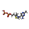
| Molecular weight | Theoretical: 425.314 Da |
| Chemical component information |  ChemComp-TPP: |
-Experimental details
-Structure determination
| Method | cryo EM |
|---|---|
 Processing Processing | single particle reconstruction |
| Aggregation state | particle |
- Sample preparation
| Concentration | 1 mg/mL | ||||||||||||
|---|---|---|---|---|---|---|---|---|---|---|---|---|---|
| Buffer | pH: 7.4 Component:
Details: 50 mM HEPES, 150 mM NaCl, 0.25 mM MgCl2 | ||||||||||||
| Grid | Model: UltrAuFoil R1.2/1.3 / Material: GOLD / Mesh: 300 / Support film - Material: GOLD / Support film - topology: HOLEY / Support film - Film thickness: 50 / Pretreatment - Type: GLOW DISCHARGE / Pretreatment - Time: 50 sec. / Pretreatment - Atmosphere: AIR / Pretreatment - Pressure: 0.04 kPa | ||||||||||||
| Vitrification | Cryogen name: ETHANE / Chamber humidity: 100 % / Chamber temperature: 277.15 K / Instrument: FEI VITROBOT MARK IV |
- Electron microscopy
| Microscope | FEI TITAN KRIOS |
|---|---|
| Specialist optics | Energy filter - Name: TFS Selectris X / Energy filter - Slit width: 10 eV |
| Software | Name: EPU |
| Image recording | Film or detector model: FEI FALCON IV (4k x 4k) / Number grids imaged: 1 / Number real images: 8045 / Average exposure time: 3.1 sec. / Average electron dose: 40.0 e/Å2 |
| Electron beam | Acceleration voltage: 300 kV / Electron source:  FIELD EMISSION GUN FIELD EMISSION GUN |
| Electron optics | Illumination mode: FLOOD BEAM / Imaging mode: BRIGHT FIELD / Nominal defocus max: 1.8 µm / Nominal defocus min: 0.6 µm |
| Sample stage | Specimen holder model: FEI TITAN KRIOS AUTOGRID HOLDER |
| Experimental equipment |  Model: Titan Krios / Image courtesy: FEI Company |
+ Image processing
-Atomic model buiding 1
| Initial model | Chain - Source name: AlphaFold / Chain - Initial model type: in silico model |
|---|---|
| Software | Name:  Coot Coot |
| Output model |  PDB-9cd3: |
 Movie
Movie Controller


 Z (Sec.)
 Y (Row.)
 X (Col.)


