 Movie
Movie Controller

[English] 日本語
 Yorodumi
- EMDB-45222: In-cell Mus musculus nuclear pore complex nuclear ring focused re... -
+ Open data

- Basic information
| Entry |  | |||||||||||||||
|---|---|---|---|---|---|---|---|---|---|---|---|---|---|---|---|---|
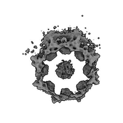
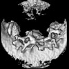
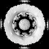
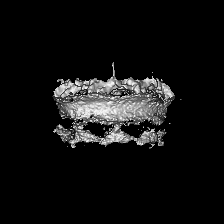
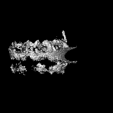
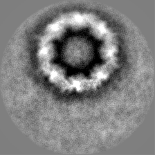
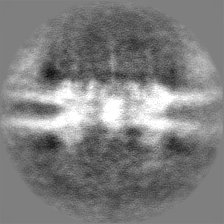
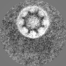
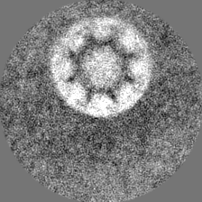
| Title | In-cell Mus musculus nuclear pore complex nuclear ring focused refinement | |||||||||||||||
 Map data Map data | In-cell Mus musculus nuclear pore complex nuclear ring focused refinement | |||||||||||||||
 Sample Sample |
| |||||||||||||||
 Keywords Keywords | Nuclear envelope / Nuclear pore / Nucleocytoplasmic transport / STRUCTURAL PROTEIN | |||||||||||||||
| Biological species |  | |||||||||||||||
| Method | subtomogram averaging / cryo EM / Resolution: 32.0 Å | |||||||||||||||
 Authors Authors | Hutchings J / Singh D / Villa E | |||||||||||||||
| Funding support |  United States, European Union, 4 items United States, European Union, 4 items
| |||||||||||||||
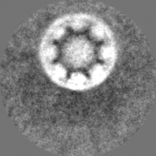
 Citation Citation | Journal: Cell / Year: 2024 Title: The molecular architecture of the nuclear basket. Authors: Digvijay Singh / Neelesh Soni / Joshua Hutchings / Ignacia Echeverria / Farhaz Shaikh / Madeleine Duquette / Sergey Suslov / Zhixun Li / Trevor van Eeuwen / Kelly Molloy / Yi Shi / Junjie ...Authors: Digvijay Singh / Neelesh Soni / Joshua Hutchings / Ignacia Echeverria / Farhaz Shaikh / Madeleine Duquette / Sergey Suslov / Zhixun Li / Trevor van Eeuwen / Kelly Molloy / Yi Shi / Junjie Wang / Qiang Guo / Brian T Chait / Javier Fernandez-Martinez / Michael P Rout / Andrej Sali / Elizabeth Villa /   Abstract: The nuclear pore complex (NPC) is the sole mediator of nucleocytoplasmic transport. Despite great advances in understanding its conserved core architecture, the peripheral regions can exhibit ...The nuclear pore complex (NPC) is the sole mediator of nucleocytoplasmic transport. Despite great advances in understanding its conserved core architecture, the peripheral regions can exhibit considerable variation within and between species. One such structure is the cage-like nuclear basket. Despite its crucial roles in mRNA surveillance and chromatin organization, an architectural understanding has remained elusive. Using in-cell cryo-electron tomography and subtomogram analysis, we explored the NPC's structural variations and the nuclear basket across fungi (yeast; S. cerevisiae), mammals (mouse; M. musculus), and protozoa (T. gondii). Using integrative structural modeling, we computed a model of the basket in yeast and mammals that revealed how a hub of nucleoporins (Nups) in the nuclear ring binds to basket-forming Mlp/Tpr proteins: the coiled-coil domains of Mlp/Tpr form the struts of the basket, while their unstructured termini constitute the basket distal densities, which potentially serve as a docking site for mRNA preprocessing before nucleocytoplasmic transport. | |||||||||||||||
| History |
|
- Structure visualization
| Supplemental images |
|---|
- Downloads & links
-EMDB archive
| Map data | emd_45222.map.gz | 39.1 MB |  EMDB map data format EMDB map data format | |
|---|---|---|---|---|
| Header (meta data) | emd-45222-v30.xmlemd-45222.xml | 13.9 KB 13.9 KB | Display Display | EMDB header |
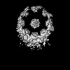
| Images |  emd_45222.png emd_45222.png | 63.5 KB | ||
| Masks | emd_45222_msk_1.map | 42.9 MB | Mask map | |
| Filedesc metadata | emd-45222.cif.gz | 4.2 KB | ||
| Others | emd_45222_half_map_1.map.gzemd_45222_half_map_2.map.gz | 33 MB 32.9 MB | ||
| Archive directory |  http://ftp.pdbj.org/pub/emdb/structures/EMD-45222ftp://ftp.pdbj.org/pub/emdb/structures/EMD-45222 http://ftp.pdbj.org/pub/emdb/structures/EMD-45222ftp://ftp.pdbj.org/pub/emdb/structures/EMD-45222 | HTTPS FTP |
-Related structure data
| Related structure data | C: citing same article ( |
|---|


-Links
| EMDB pages | EMDB (EBI/PDBe) / EMDataResource |
|---|
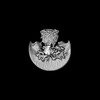
-Map
| File | Download / File: emd_45222.map.gz / Format: CCP4 / Size: 42.9 MB / Type: IMAGE STORED AS FLOATING POINT NUMBER (4 BYTES) | ||||||||||||||||||||||||||||||||||||
|---|---|---|---|---|---|---|---|---|---|---|---|---|---|---|---|---|---|---|---|---|---|---|---|---|---|---|---|---|---|---|---|---|---|---|---|---|---|
| Annotation | In-cell Mus musculus nuclear pore complex nuclear ring focused refinement | ||||||||||||||||||||||||||||||||||||
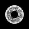
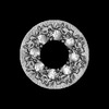
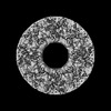
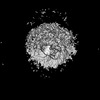
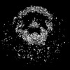
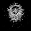
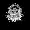
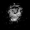
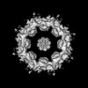
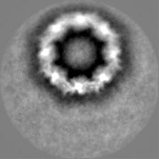
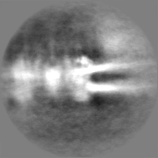
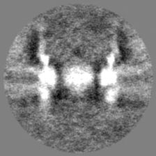
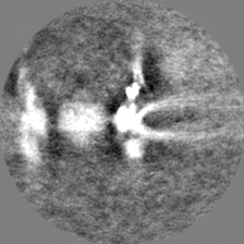
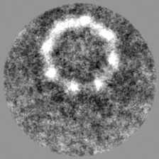
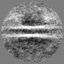
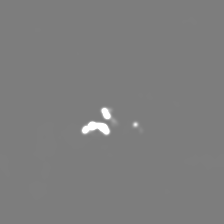
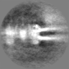
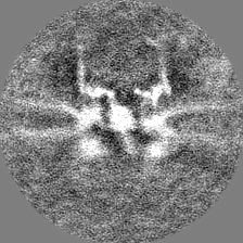
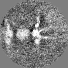
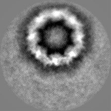
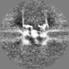
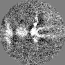
| Projections & slices | Image control
Images are generated by Spider. | ||||||||||||||||||||||||||||||||||||
| Voxel size | X=Y=Z: 10 Å | ||||||||||||||||||||||||||||||||||||
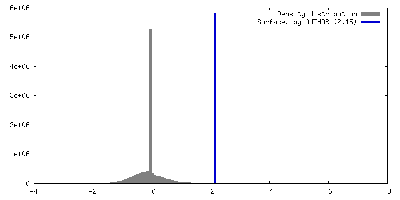
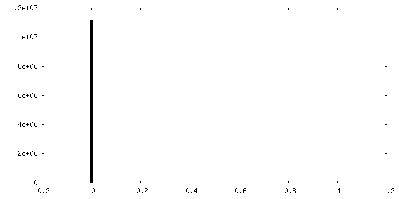
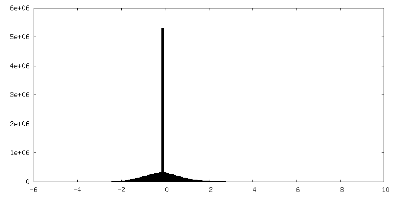
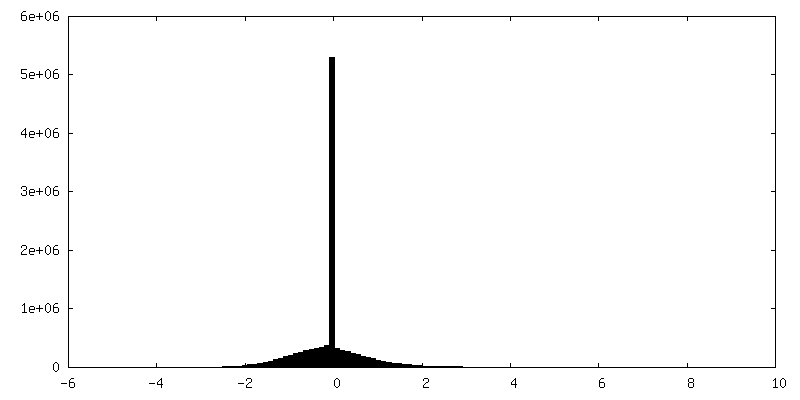
| Density |
| ||||||||||||||||||||||||||||||||||||
| Symmetry | Space group: 1 | ||||||||||||||||||||||||||||||||||||
| Details | EMDB XML:
|
 Z (Sec.)
 Y (Row.)
 X (Col.)


-Supplemental data
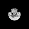
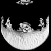
-Mask #1
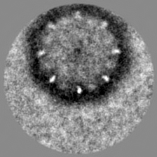
| File | emd_45222_msk_1.map | ||||||||||||
|---|---|---|---|---|---|---|---|---|---|---|---|---|---|
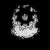
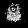
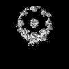
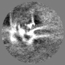
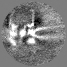
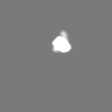
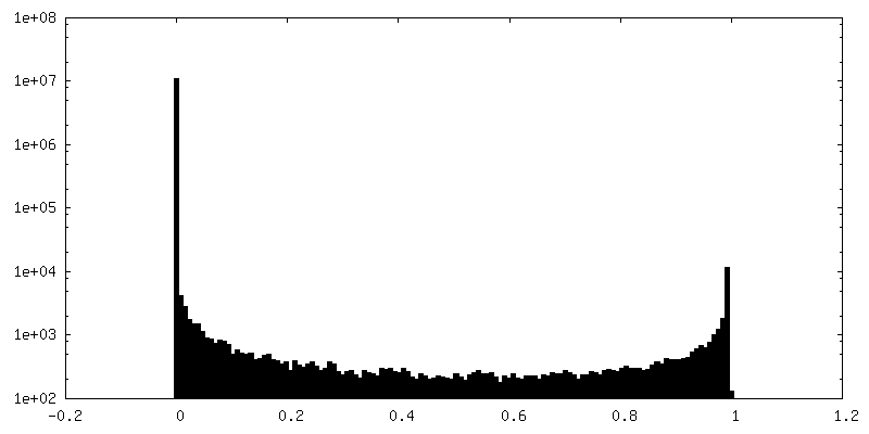
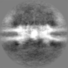
| Projections & Slices |
| ||||||||||||
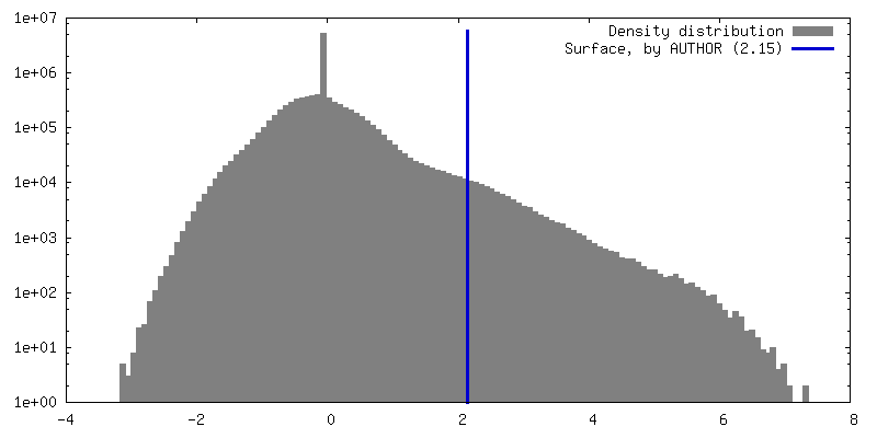
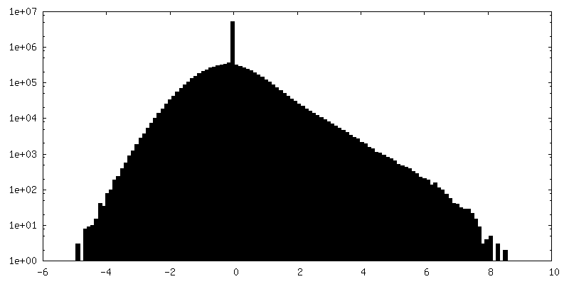
| Density Histograms |


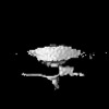
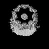
-Half map: In-cell Mus musculus nuclear pore complex nuclear ring...
| File | emd_45222_half_map_1.map | ||||||||||||
|---|---|---|---|---|---|---|---|---|---|---|---|---|---|
| Annotation | In-cell Mus musculus nuclear pore complex nuclear ring focused refinement - half map 1 | ||||||||||||
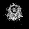
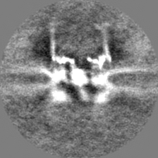
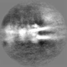
| Projections & Slices |
| ||||||||||||
| Density Histograms |

-Half map: In-cell Mus musculus nuclear pore complex nuclear ring...
| File | emd_45222_half_map_2.map | ||||||||||||
|---|---|---|---|---|---|---|---|---|---|---|---|---|---|
| Annotation | In-cell Mus musculus nuclear pore complex nuclear ring focused refinement - half map 1 | ||||||||||||
| Projections & Slices |
| ||||||||||||
| Density Histograms |
- Sample components
-Entire : In-cell Mus musculus nuclear pore complex nuclear ring focused re...
| Entire | Name: In-cell Mus musculus nuclear pore complex nuclear ring focused refinement |
|---|---|
| Components |
|
-Supramolecule #1: In-cell Mus musculus nuclear pore complex nuclear ring focused re...
| Supramolecule | Name: In-cell Mus musculus nuclear pore complex nuclear ring focused refinement type: cell / ID: 1 / Parent: 0 |
|---|---|
| Source (natural) | Organism: |
-Experimental details
-Structure determination
| Method | cryo EM |
|---|---|
 Processing Processing | subtomogram averaging |
| Aggregation state | cell |
-Sample preparation
| Buffer | pH: 7.4 |
|---|---|
| Vitrification | Cryogen name: ETHANE-PROPANE / Instrument: HOMEMADE PLUNGER |
- Electron microscopy
| Microscope | FEI TITAN KRIOS |
|---|---|
| Image recording | Film or detector model: GATAN K3 BIOCONTINUUM (6k x 4k) / Detector mode: COUNTING / Digitization - Dimensions - Width: 5760 pixel / Digitization - Dimensions - Height: 4092 pixel / Average electron dose: 3.7 e/Å2 |
| Electron beam | Acceleration voltage: 300 kV / Electron source:  FIELD EMISSION GUN FIELD EMISSION GUN |
| Electron optics | Illumination mode: FLOOD BEAM / Imaging mode: BRIGHT FIELD / Nominal defocus max: 5.0 µm / Nominal defocus min: 3.0 µm |
| Experimental equipment |  Model: Titan Krios / Image courtesy: FEI Company |
-Image processing
| Final reconstruction | Applied symmetry - Point group: C1 (asymmetric) / Resolution.type: BY AUTHOR / Resolution: 32.0 Å / Resolution method: FSC 0.143 CUT-OFF / Number subtomograms used: 806 |
|---|---|
| Extraction | Number tomograms: 136 / Number images used: 220 |
| Final angle assignment | Type: MAXIMUM LIKELIHOOD |
