[English] 日本語
Yorodumi
- EMDB-45151: Hexadecamer of NL4-3 WT HIV-1 intasome -

+
Open data


ID or keywords:

Loading...

-
Basic information

Entry
Database: EMDB / ID: EMD-45151
TitleHexadecamer of NL4-3 WT HIV-1 intasome
Map dataThis is a composite map
Sample
  • Complex: crosslinked wild-type NL4-3 HIV-1 Intasome
    • Protein or peptide: Integrase
    • DNA: DNA (5'-D(*AP*CP*TP*GP*CP*TP*AP*GP*AP*GP*AP*TP*TP*TP*TP*CP*CP*CP*G)-3')
    • DNA: DNA (5'-D(P*CP*GP*GP*GP*AP*AP*AP*AP*TP*CP*TP*CP*TP*AP*GP*CP*A)-3')
  • Ligand: MAGNESIUM ION
KeywordsViral protein / protein complex / VIRAL PROTEIN-DNA complex
Function / homology
Function and homology information


HIV-1 retropepsin / symbiont-mediated activation of host apoptosis / retroviral ribonuclease H / exoribonuclease H / exoribonuclease H activity / host multivesicular body / DNA integration / viral genome integration into host DNA / RNA-directed DNA polymerase / establishment of integrated proviral latency ...HIV-1 retropepsin / symbiont-mediated activation of host apoptosis / retroviral ribonuclease H / exoribonuclease H / exoribonuclease H activity / host multivesicular body / DNA integration / viral genome integration into host DNA / RNA-directed DNA polymerase / establishment of integrated proviral latency / telomerase activity / viral penetration into host nucleus / RNA stem-loop binding / RNA-DNA hybrid ribonuclease activity / Transferases; Transferring phosphorus-containing groups; Nucleotidyltransferases / host cell / viral nucleocapsid / DNA recombination / DNA-directed DNA polymerase / aspartic-type endopeptidase activity / Hydrolases; Acting on ester bonds / DNA-directed DNA polymerase activity / symbiont-mediated suppression of host gene expression / symbiont entry into host cell / viral translational frameshifting / lipid binding / host cell nucleus / host cell plasma membrane / virion membrane / structural molecule activity / proteolysis / DNA binding / zinc ion binding / membrane
Similarity search - Function
Reverse transcriptase connection / Reverse transcriptase connection domain / Reverse transcriptase thumb / Reverse transcriptase thumb domain / Integrase Zinc binding domain / Zinc finger integrase-type profile. / Integrase-like, N-terminal / Integrase DNA binding domain / Integrase, C-terminal domain superfamily, retroviral / Integrase, N-terminal zinc-binding domain ...Reverse transcriptase connection / Reverse transcriptase connection domain / Reverse transcriptase thumb / Reverse transcriptase thumb domain / Integrase Zinc binding domain / Zinc finger integrase-type profile. / Integrase-like, N-terminal / Integrase DNA binding domain / Integrase, C-terminal domain superfamily, retroviral / Integrase, N-terminal zinc-binding domain / Integrase, C-terminal, retroviral / Integrase DNA binding domain profile. / Immunodeficiency lentiviral matrix, N-terminal / gag gene protein p17 (matrix protein) / RNase H / Integrase core domain / Integrase, catalytic core / Integrase catalytic domain profile. / Retropepsin-like catalytic domain / Matrix protein, lentiviral and alpha-retroviral, N-terminal / Retroviral nucleocapsid Gag protein p24, C-terminal domain / Gag protein p24 C-terminal domain / RNase H type-1 domain profile. / Ribonuclease H domain / Retropepsins / Retroviral aspartyl protease / Aspartyl protease, retroviral-type family profile. / Peptidase A2A, retrovirus, catalytic / Reverse transcriptase domain / Reverse transcriptase (RNA-dependent DNA polymerase) / Reverse transcriptase (RT) catalytic domain profile. / Retrovirus capsid, C-terminal / Retroviral matrix protein / Retrovirus capsid, N-terminal / zinc finger / Zinc knuckle / Zinc finger, CCHC-type superfamily / Zinc finger, CCHC-type / Zinc finger CCHC-type profile. / Aspartic peptidase, active site / Eukaryotic and viral aspartyl proteases active site. / Aspartic peptidase domain superfamily / Ribonuclease H superfamily / Ribonuclease H-like superfamily / Reverse transcriptase/Diguanylate cyclase domain / DNA/RNA polymerase superfamily
Similarity search - Domain/homology
Biological speciesHIV type 1 (virus) / HIV-1 06TG.HT008 (virus)
Methodsingle particle reconstruction / cryo EM / Resolution: 8.0 Å
AuthorsLyumkis D / Jing T / Zhang Z / Biswas A
Funding support United States, 5 items
OrganizationGrant numberCountry
National Institutes of Health/National Institute Of Allergy and Infectious Diseases (NIH/NIAID)AI136680 United States
National Institutes of Health/National Institute Of Allergy and Infectious Diseases (NIH/NIAID)AI146017 United States
National Institutes of Health/National Institute Of Allergy and Infectious Diseases (NIH/NIAID)AI170855 United States
National Institutes of Health/National Institute Of Allergy and Infectious Diseases (NIH/NIAID)AI039394 United States
National Institutes of Health/National Institute Of Allergy and Infectious Diseases (NIH/NIAID)AI170791 United States
CitationJournal: Biorxiv / Year: 2025
Title: Oligomeric HIV-1 Integrase Structures Reveal Functional Plasticity for Intasome Assembly and RNA Binding
Authors: Jing T / Shan Z / Dinh T / Biswas A / Jang S / Greenwood J / Li M / Zhang Z / Gray G / Shin HJ / Zhou B / Passos D / Strutzenberg TS / Aiyer S / Andrade L / Zhang Y / Li Z / Craigie R / ...Authors: Jing T / Shan Z / Dinh T / Biswas A / Jang S / Greenwood J / Li M / Zhang Z / Gray G / Shin HJ / Zhou B / Passos D / Strutzenberg TS / Aiyer S / Andrade L / Zhang Y / Li Z / Craigie R / Engelman AN / Kvaratskhelia M / Lyumkis D
History
DepositionMay 30, 2024-
Header (metadata) releaseJun 25, 2025-
Map releaseJun 25, 2025-
UpdateJun 25, 2025-
Current statusJun 25, 2025Processing site: RCSB / Status: Released

-
Structure visualization

Supplemental images

Downloads & links

-
Map

FileDownload / File: emd_45151.map.gz / Format: CCP4 / Size: 125 MB / Type: IMAGE STORED AS FLOATING POINT NUMBER (4 BYTES)
AnnotationThis is a composite map
Projections & slices

Image control

Size
Brightness
Contrast
Others
AxesZ (Sec.)Y (Row.)X (Col.)
1.02 Å/pix.
x 320 pix.
= 324.8 Å
1.02 Å/pix.
x 320 pix.
= 324.8 Å
1.02 Å/pix.
x 320 pix.
= 324.8 Å

Surface

Projections

Slices (1/3)

Slices (1/2)

Slices (2/3)

Images are generated by Spider.

Voxel sizeX=Y=Z: 1.015 Å
Density
Contour LevelBy AUTHOR: 0.4
Minimum - Maximum-0.28248605 - 1.8204563
Average (Standard dev.)0.014856143 (±0.0937968)
SymmetrySpace group: 1
Details

EMDB XML:

Map geometry
Axis orderXYZ
Origin000
Dimensions320320320
Spacing320320320
CellA=B=C: 324.8 Å
α=β=γ: 90.0 °

-
Supplemental data

-
Mask #1

Fileemd_45151_msk_1.map
Projections & Slices
AxesZYX

Projections

Slices (1/2)
Density Histograms

-
Sample components

-
Entire : crosslinked wild-type NL4-3 HIV-1 Intasome

EntireName: crosslinked wild-type NL4-3 HIV-1 Intasome
Components
  • Complex: crosslinked wild-type NL4-3 HIV-1 Intasome
    • Protein or peptide: Integrase
    • DNA: DNA (5'-D(*AP*CP*TP*GP*CP*TP*AP*GP*AP*GP*AP*TP*TP*TP*TP*CP*CP*CP*G)-3')
    • DNA: DNA (5'-D(P*CP*GP*GP*GP*AP*AP*AP*AP*TP*CP*TP*CP*TP*AP*GP*CP*A)-3')
  • Ligand: MAGNESIUM ION

-
Supramolecule #1: crosslinked wild-type NL4-3 HIV-1 Intasome

SupramoleculeName: crosslinked wild-type NL4-3 HIV-1 Intasome / type: complex / ID: 1 / Parent: 0 / Macromolecule list: #1-#3
Source (natural)Organism: HIV type 1 (virus)
Molecular weightTheoretical: 600 KDa

-
Macromolecule #1: Integrase

MacromoleculeName: Integrase / type: protein_or_peptide / ID: 1 / Number of copies: 16 / Enantiomer: LEVO
EC number: Transferases; Transferring phosphorus-containing groups; Nucleotidyltransferases
Source (natural)Organism: HIV-1 06TG.HT008 (virus)
Molecular weightTheoretical: 32.244787 KDa
Recombinant expressionOrganism: Escherichia coli 'BL21-Gold(DE3)pLysS AG' (bacteria)
SequenceString: FLDGIDKAQE EHEKYHSNWR AMASDFNLPP VVAKEIVASC DKCQLKGEAM HGQVDCSPGI WQLDCTHLEG KVILVAVHVA SGYIEAEVI PAETGQETAY FLLKLAGRWP VKTVHTDNGS NFTSTTVKAA CWWAGIKQEF GIPYNPQSQG VIESMNKELK K IIGQVRDQ ...String:
FLDGIDKAQE EHEKYHSNWR AMASDFNLPP VVAKEIVASC DKCQLKGEAM HGQVDCSPGI WQLDCTHLEG KVILVAVHVA SGYIEAEVI PAETGQETAY FLLKLAGRWP VKTVHTDNGS NFTSTTVKAA CWWAGIKQEF GIPYNPQSQG VIESMNKELK K IIGQVRDQ AEHLKTAVQM AVFIHNFKRK GGIGGYSAGE RIVDIIATDI QTKELQKQIT KIQNFRVYYR DSRDPVWKGP AK LLWKGEG AVVIQDNSDI KVVPRRKAKI IRDYGKQMAG DDCVASRQDE D

UniProtKB: Gag-Pol polyprotein

-
Macromolecule #2: DNA (5'-D(*AP*CP*TP*GP*CP*TP*AP*GP*AP*GP*AP*TP*TP*TP*TP*CP*CP*CP*...

MacromoleculeName: DNA (5'-D(*AP*CP*TP*GP*CP*TP*AP*GP*AP*GP*AP*TP*TP*TP*TP*CP*CP*CP*G)-3')
type: dna / ID: 2 / Number of copies: 2 / Classification: DNA
Source (natural)Organism: HIV-1 06TG.HT008 (virus)
Molecular weightTheoretical: 5.795758 KDa
SequenceString:
(DA)(DC)(DT)(DG)(DC)(DT)(DA)(DG)(DA)(DG) (DA)(DT)(DT)(DT)(DT)(DC)(DC)(DC)(DG)

-
Macromolecule #3: DNA (5'-D(P*CP*GP*GP*GP*AP*AP*AP*AP*TP*CP*TP*CP*TP*AP*GP*CP*A)-3')

MacromoleculeName: DNA (5'-D(P*CP*GP*GP*GP*AP*AP*AP*AP*TP*CP*TP*CP*TP*AP*GP*CP*A)-3')
type: dna / ID: 3 / Number of copies: 2 / Classification: DNA
Source (natural)Organism: HIV-1 06TG.HT008 (virus)
Molecular weightTheoretical: 5.220413 KDa
SequenceString:
(DC)(DG)(DG)(DG)(DA)(DA)(DA)(DA)(DT)(DC) (DT)(DC)(DT)(DA)(DG)(DC)(DA)

-
Macromolecule #4: MAGNESIUM ION

MacromoleculeName: MAGNESIUM ION / type: ligand / ID: 4 / Number of copies: 4 / Formula: MG
Molecular weightTheoretical: 24.305 Da

-
Experimental details

-
Structure determination

Methodcryo EM
Processingsingle particle reconstruction
Aggregation stateparticle

-
Sample preparation

BufferpH: 6.2
Component:
ConcentrationNameFormula
20.0 mMTris-HCl
1000.0 mMsodium chlorideNaCl
0.5 mMTCEP
10.0 %glycerolC3H5(OH)3
5.0 mMMagnesium chlorideMgCl2
GridModel: Quantifoil R1.2/1.3 / Material: GOLD / Support film - Material: GOLD / Support film - topology: HOLEY
VitrificationCryogen name: ETHANE / Instrument: HOMEMADE PLUNGER
Details: Cryo-EM grids were prepared by freezing using a manual plunger in cold room at 4C.

-
Electron microscopy

MicroscopeTFS KRIOS
Image recordingFilm or detector model: GATAN K2 SUMMIT (4k x 4k) / Detector mode: COUNTING / Digitization - Dimensions - Width: 3710 pixel / Digitization - Dimensions - Height: 3838 pixel / Digitization - Frames/image: 1-50 / Number real images: 775 / Average exposure time: 12.0 sec. / Average electron dose: 60.0 e/Å2
Electron beamAcceleration voltage: 300 kV / Electron source: FIELD EMISSION GUN
Electron opticsC2 aperture diameter: 50.0 µm / Illumination mode: FLOOD BEAM / Imaging mode: BRIGHT FIELD / Cs: 2.7 mm / Nominal defocus max: 2.2 µm / Nominal defocus min: 0.8 µm / Nominal magnification: 29000
Sample stageSpecimen holder model: FEI TITAN KRIOS AUTOGRID HOLDER / Cooling holder cryogen: NITROGEN
Experimental equipment
Model: Titan Krios / Image courtesy: FEI Company

+
Image processing

CTF correctionSoftware - Name: cryoSPARC (ver. 4.2) / Type: PHASE FLIPPING AND AMPLITUDE CORRECTION
Startup modelType of model: OTHER
Final reconstructionApplied symmetry - Point group: C2 (2 fold cyclic) / Resolution.type: BY AUTHOR / Resolution: 8.0 Å / Resolution method: OTHER / Software - Name: cryoSPARC (ver. 4.2)
Details: 3D FSC server is used to compute map-model resolution at the threshold of 0.5. This reported resolution is not the map resolution because it is a composite map that does not have two half maps.
Number images used: 22780
Initial angle assignmentType: PROJECTION MATCHING / Software - Name: cryoSPARC (ver. 4.2)
Final angle assignmentType: PROJECTION MATCHING / Software - Name: cryoSPARC (ver. 4.2)

-
Atomic model buiding 1

Initial model
PDB IDChain

source_name: PDB, initial_model_type: experimental model

source_name: PDB, initial_model_type: experimental model

source_name: PDB, initial_model_type: experimental model

source_name: PDB, initial_model_type: experimental model

source_name: PDB, initial_model_type: experimental model
Details
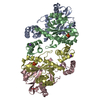
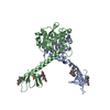
Domains of HIV-1 IN were rigid-body docked into cryo-EM density without any further refinement or adjustment of the coordinates. This rigid-body docked model was then truncated to poly-Alanine and deposited here.
RefinementSpace: REAL / Protocol: RIGID BODY FIT / Target criteria: Correlation coefficient
Output model

PDB-9c29:
Hexadecamer of NL4-3 WT HIV-1 intasome

+
About Yorodumi

-
News

-
Feb 9, 2022. New format data for meta-information of EMDB entries

New format data for meta-information of EMDB entries

  • Version 3 of the EMDB header file is now the official format.
  • The previous official version 1.9 will be removed from the archive.

Related info.:EMDB header

External links:wwPDB to switch to version 3 of the EMDB data model

-
Aug 12, 2020. Covid-19 info

Covid-19 info

URL: https://pdbj.org/emnavi/covid19.php

New page: Covid-19 featured information page in EM Navigator.

Related info.:Covid-19 info / Mar 5, 2020. Novel coronavirus structure data

+
Mar 5, 2020. Novel coronavirus structure data

Novel coronavirus structure data

Related info.:Yorodumi Speices / Aug 12, 2020. Covid-19 info

External links:COVID-19 featured content - PDBj / Molecule of the Month (242):Coronavirus Proteases

+
Jan 31, 2019. EMDB accession codes are about to change! (news from PDBe EMDB page)

EMDB accession codes are about to change! (news from PDBe EMDB page)

  • The allocation of 4 digits for EMDB accession codes will soon come to an end. Whilst these codes will remain in use, new EMDB accession codes will include an additional digit and will expand incrementally as the available range of codes is exhausted. The current 4-digit format prefixed with “EMD-” (i.e. EMD-XXXX) will advance to a 5-digit format (i.e. EMD-XXXXX), and so on. It is currently estimated that the 4-digit codes will be depleted around Spring 2019, at which point the 5-digit format will come into force.
  • The EM Navigator/Yorodumi systems omit the EMD- prefix.

Related info.:Q: What is EMD? / ID/Accession-code notation in Yorodumi/EM Navigator

External links:EMDB Accession Codes are Changing Soon! / Contact to PDBj

+
Jul 12, 2017. Major update of PDB

Major update of PDB

  • wwPDB released updated PDB data conforming to the new PDBx/mmCIF dictionary.
  • This is a major update changing the version number from 4 to 5, and with Remediation, in which all the entries are updated.
  • In this update, many items about electron microscopy experimental information are reorganized (e.g. em_software).
  • Now, EM Navigator and Yorodumi are based on the updated data.

External links:wwPDB Remediation / Enriched Model Files Conforming to OneDep Data Standards Now Available in the PDB FTP Archive

-
Yorodumi

Thousand views of thousand structures

  • Yorodumi is a browser for structure data from EMDB, PDB, SASBDB, etc.
  • This page is also the successor to EM Navigator detail page, and also detail information page/front-end page for Omokage search.
  • The word "yorodu" (or yorozu) is an old Japanese word meaning "ten thousand". "mi" (miru) is to see.

Related info.:EMDB / PDB / SASBDB / Comparison of 3 databanks / Yorodumi Search / Aug 31, 2016. New EM Navigator & Yorodumi / Yorodumi Papers / Jmol/JSmol / Function and homology information / Changes in new EM Navigator and Yorodumi

Read more