 Movie
Movie Controller


+ Open data

- Basic information
| Entry | 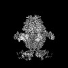 | |||||||||
|---|---|---|---|---|---|---|---|---|---|---|
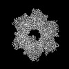
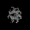
| Title | HerA-DUF assembly 1 | |||||||||
 Map data Map data | DeepEmhancer refined map | |||||||||
 Sample Sample |
| |||||||||
 Keywords Keywords | anti-phage / nuclease / ATPase / 18-mer / ANTIVIRAL PROTEIN | |||||||||
| Function / homology | Helicase HerA-like / Helicase HerA, central domain / Helicase HerA, central domain / P-loop containing nucleoside triphosphate hydrolase / ATP binding / ATP-binding protein / DUF4297 domain-containing protein Function and homology information Function and homology information | |||||||||
| Biological species |  | |||||||||
| Method | single particle reconstruction / cryo EM / Resolution: 2.76 Å | |||||||||
 Authors Authors | Rish AD / Fosuah E / Fu TM | |||||||||
| Funding support |  United States, 1 items United States, 1 items
| |||||||||
 Citation Citation | Journal: Mol Cell / Year: 2025 Title: Architecture remodeling activates the HerA-DUF anti-phage defense system. Authors: Anthony D Rish / Elizabeth Fosuah / Zhangfei Shen / Ila A Marathe / Vicki H Wysocki / Tian-Min Fu / Abstract: Leveraging AlphaFold models and integrated experiments, we characterized the HerA-DUF4297 (DUF) anti-phage defense system, focusing on DUF's undefined biochemical functions. Guided by structure-based ...Leveraging AlphaFold models and integrated experiments, we characterized the HerA-DUF4297 (DUF) anti-phage defense system, focusing on DUF's undefined biochemical functions. Guided by structure-based genomic analyses, we found DUF homologs to be universally distributed across diverse bacterial immune systems. Notably, one such homolog, Cap4, is a nuclease. Inspired by this evolutionary clue, we tested DUF's nuclease activity and observed that DUF cleaves DNA substrates only when bound to its partner protein HerA. To dissect the mechanism of DUF activation, we determined the structures of DUF and HerA-DUF. Although DUF forms large oligomeric assemblies both alone and with HerA, oligomerization alone was insufficient to elicit nuclease activity. Instead, HerA binding induces a profound architecture remodeling that propagates throughout the complex. This remodeling reconfigures DUF into an active nuclease capable of robust DNA cleavage. Together, we highlight an architecture remodeling-driven mechanism that may inform the activation of other immune systems. | |||||||||
| History |
|
- Structure visualization
| Supplemental images |
|---|

- Downloads & links
-EMDB archive
| Map data | emd_45124.map.gz | 126.5 MB | EMDB map data format | |
|---|---|---|---|---|
| Header (meta data) | emd-45124-v30.xmlemd-45124.xml | 20 KB 20 KB | Display Display | EMDB header |
| FSC (resolution estimation) | emd_45124_fsc.xml | 11 KB | Display | FSC data file |
| Images |  emd_45124.png emd_45124.png | 78.7 KB | ||
| Filedesc metadata | emd-45124.cif.gz | 6.2 KB | ||
| Others | emd_45124_additional_1.map.gzemd_45124_additional_2.map.gzemd_45124_half_map_1.map.gzemd_45124_half_map_2.map.gz | 136.5 MB 125.8 MB 134.3 MB 134.3 MB | ||
| Archive directory |  http://ftp.pdbj.org/pub/emdb/structures/EMD-45124ftp://ftp.pdbj.org/pub/emdb/structures/EMD-45124 http://ftp.pdbj.org/pub/emdb/structures/EMD-45124ftp://ftp.pdbj.org/pub/emdb/structures/EMD-45124 | HTTPS FTP |
-Validation report
| Summary document | emd_45124_validation.pdf.gz | 781.9 KB | Display | EMDB validaton report |
|---|---|---|---|---|
| Full document | emd_45124_full_validation.pdf.gz | 781.5 KB | Display | |
| Data in XML | emd_45124_validation.xml.gz | 19.2 KB | Display | |
| Data in CIF | emd_45124_validation.cif.gz | 25.2 KB | Display | |
| Arichive directory | https://ftp.pdbj.org/pub/emdb/validation_reports/EMD-45124ftp://ftp.pdbj.org/pub/emdb/validation_reports/EMD-45124 | HTTPS FTP |
-Related structure data
| Related structure data |  9c1mMC  9c1nC 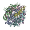 9c1oC  9c1xC  9c5xC M: atomic model generated by this map C: citing same article ( |
|---|---|
| Similar structure data |

-Links
| EMDB pages | EMDB (EBI/PDBe) / EMDataResource |
|---|
-Map
| File | Download / File: emd_45124.map.gz / Format: CCP4 / Size: 144.7 MB / Type: IMAGE STORED AS FLOATING POINT NUMBER (4 BYTES) | ||||||||||||||||||||||||||||||||||||
|---|---|---|---|---|---|---|---|---|---|---|---|---|---|---|---|---|---|---|---|---|---|---|---|---|---|---|---|---|---|---|---|---|---|---|---|---|---|
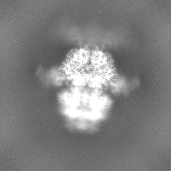
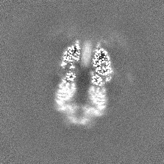
| Annotation | DeepEmhancer refined map | ||||||||||||||||||||||||||||||||||||
| Projections & slices | Image control
Images are generated by Spider. | ||||||||||||||||||||||||||||||||||||
| Voxel size | X=Y=Z: 1.09 Å | ||||||||||||||||||||||||||||||||||||
| Density |
| ||||||||||||||||||||||||||||||||||||
| Symmetry | Space group: 1 | ||||||||||||||||||||||||||||||||||||
| Details | EMDB XML:
|
 Z (Sec.)
 Y (Row.)
 X (Col.)


-Supplemental data
-Additional map: cryoSparc refined map
| File | emd_45124_additional_1.map | ||||||||||||
|---|---|---|---|---|---|---|---|---|---|---|---|---|---|
| Annotation | cryoSparc refined map | ||||||||||||
| Projections & Slices |
| ||||||||||||
| Density Histograms |


-Additional map: cryosparc refine before C6 symmetry expansion
| File | emd_45124_additional_2.map | ||||||||||||
|---|---|---|---|---|---|---|---|---|---|---|---|---|---|
| Annotation | cryosparc refine before C6 symmetry expansion | ||||||||||||
| Projections & Slices |
| ||||||||||||
| Density Histograms |


-Half map: Half map B
| File | emd_45124_half_map_1.map | ||||||||||||
|---|---|---|---|---|---|---|---|---|---|---|---|---|---|
| Annotation | Half map B | ||||||||||||
| Projections & Slices |
| ||||||||||||
| Density Histograms |


-Half map: Half map A
| File | emd_45124_half_map_2.map | ||||||||||||
|---|---|---|---|---|---|---|---|---|---|---|---|---|---|
| Annotation | Half map A | ||||||||||||
| Projections & Slices |
| ||||||||||||
| Density Histograms |


- Sample components
-Entire : HerA-DUF4297
| Entire | Name: HerA-DUF4297 |
|---|---|
| Components |
|
-Supramolecule #1: HerA-DUF4297
| Supramolecule | Name: HerA-DUF4297 / type: complex / ID: 1 / Parent: 0 / Macromolecule list: all / Details: Anti-phage defense supramolecular complex |
|---|---|
| Source (natural) | Organism: |
| Molecular weight | Theoretical: 1.02 MDa |
-Macromolecule #1: DUF4297 domain-containing protein
| Macromolecule | Name: DUF4297 domain-containing protein / type: protein_or_peptide / ID: 1 / Number of copies: 12 / Enantiomer: LEVO |
|---|---|
| Source (natural) | Organism: |
| Molecular weight | Theoretical: 51.330734 KDa |
| Recombinant expression | Organism: |
| Sequence | String: MMSREADHTI KGFLYQFNKT LNSILSSTDQ DEIQIEGIIE DIDIKNSNIT NAIQCKYHES KVRHNLSDIY KPILQMLLHF LENDSLNIK YALYAYFPNE QVGVKEVTKS QIEEILSSSN FDYISKYISK IKPPKEQIIK ELLGKTSKTT EDKTRIKKYY E TSKLETIV ...String: MMSREADHTI KGFLYQFNKT LNSILSSTDQ DEIQIEGIIE DIDIKNSNIT NAIQCKYHES KVRHNLSDIY KPILQMLLHF LENDSLNIK YALYAYFPNE QVGVKEVTKS QIEEILSSSN FDYISKYISK IKPPKEQIIK ELLGKTSKTT EDKTRIKKYY E TSKLETIV DIDKFLRDHF VFEIGLSYEE LMNETKNLLM KEGFSLEDVK DLFYPNSIQY IAELSILPEA EKRISSKNKL ID YLKGNKK TAMSRWTSEV LTRKQLLKVR KNQLVPSLNI NSRSRYFIID PDTIDNFDDE FILFVKDYLD KYNSKIKLHT ETP CFILKT DVNNLSEYHK RFVSRNIQII TGYIGDTFYF KEFNKEPKRI IKDNWVEFKA RISCNSDEVI KCINYKKCDD LYIV GGVDV SLLDTADVNI ENLEINNFRE LKYLLSMLKE I UniProtKB: DUF4297 domain-containing protein |
-Macromolecule #2: ATP-binding protein
| Macromolecule | Name: ATP-binding protein / type: protein_or_peptide / ID: 2 / Number of copies: 6 / Enantiomer: LEVO |
|---|---|
| Source (natural) | Organism: |
| Molecular weight | Theoretical: 67.182125 KDa |
| Recombinant expression | Organism: |
| Sequence | String: MKIGSVIESS PHSILVKIDT LKIFEKAKSA LQIGKYLKIQ EGNHNFVLCV IQNIKISTDK DEDIFILTVQ PVGIFKGEEF FQGNSMLPS PTEPVFLVED DILNKIFSNE KTKIFHLGNL AQNEEVSFTL DGDKFFSKHV AVVGSTGSGK SCAVAKILQN V VGINDARN ...String: MKIGSVIESS PHSILVKIDT LKIFEKAKSA LQIGKYLKIQ EGNHNFVLCV IQNIKISTDK DEDIFILTVQ PVGIFKGEEF FQGNSMLPS PTEPVFLVED DILNKIFSNE KTKIFHLGNL AQNEEVSFTL DGDKFFSKHV AVVGSTGSGK SCAVAKILQN V VGINDARN INKSDKKNSH IIIFDIHSEY KSAFEIDKNE DFNLNYLDVE KLKLPYWLMN SEELETLFIE SNEQNSHNQV SQ FKRAVVL NKEKYNPEFK KITYDSPVYF NINEVFNYIY NLNEEVINKI EGEPSLPKLS NGELVENRQI YFNEKLEFTS SNT SKATKA SNGPFNGEFN RFLSRFETKL TDKRLEFLLL NQDVEENSKY RTEHFEDILK QFMGYLDRSN VSIIDLSGIP FEVL SITIS LISRLIFDFA FHYSKLQHQK DELNDIPFMI VCEEAHNYIP RTGGIEFKAA KKSIERIAKE GRKYGLSLMV VSQRP SEVS DTILSQCNNF INLRLTNIND QNYIKNLLPD NSRSISEILP TLGAGECLVV GDSTPIPSIV KLELPNPEPR SQSIKF HKK WSESWRTPSF EEVIMRWRKE NG UniProtKB: ATP-binding protein |
-Experimental details
-Structure determination
| Method | cryo EM |
|---|---|
 Processing Processing | single particle reconstruction |
| Aggregation state | particle |
-Sample preparation
| Concentration | 1.75 mg/mL |
|---|---|
| Buffer | pH: 7.5 |
| Vitrification | Cryogen name: ETHANE |
- Electron microscopy
| Microscope | FEI TITAN KRIOS |
|---|---|
| Image recording | Film or detector model: GATAN K3 BIOQUANTUM (6k x 4k) / Average electron dose: 50.0 e/Å2 |
| Electron beam | Acceleration voltage: 300 kV / Electron source:  FIELD EMISSION GUN FIELD EMISSION GUN |
| Electron optics | Illumination mode: FLOOD BEAM / Imaging mode: BRIGHT FIELD / Nominal defocus max: 2.0 µm / Nominal defocus min: 0.5 µm |
| Experimental equipment |  Model: Titan Krios / Image courtesy: FEI Company |

