[English] 日本語
 Yorodumi
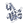
- EMDB-44902: Cryo-EM Structure of AAV2 Rep68 bound to integration site AAVS1 -
+ Open data
- Basic information
| Entry |  | |||||||||
|---|---|---|---|---|---|---|---|---|---|---|
| Title | Cryo-EM Structure of AAV2 Rep68 bound to integration site AAVS1 | |||||||||
 Map data Map data | ||||||||||
 Sample Sample |
| |||||||||
 Keywords Keywords | AAV / Protein-DNA complex / Replication / Helicase / AAA+ / SF3 / DNA BINDING PROTEIN / VIRAL PROTEIN-DNA complex | |||||||||
| Function / homology |  Function and homology information Function and homology informationsymbiont-mediated arrest of host cell cycle during G2/M transition / symbiont entry into host cell via permeabilization of host membrane / viral DNA genome replication / symbiont-mediated perturbation of host cell cycle G1/S transition checkpoint / endonuclease activity / DNA helicase / DNA replication / host cell nucleus / ATP hydrolysis activity / DNA binding ...symbiont-mediated arrest of host cell cycle during G2/M transition / symbiont entry into host cell via permeabilization of host membrane / viral DNA genome replication / symbiont-mediated perturbation of host cell cycle G1/S transition checkpoint / endonuclease activity / DNA helicase / DNA replication / host cell nucleus / ATP hydrolysis activity / DNA binding / ATP binding / metal ion binding Similarity search - Function | |||||||||
| Biological species |  adeno-associated virus 2 / adeno-associated virus 2 /  Homo sapiens (human) Homo sapiens (human) | |||||||||
| Method | single particle reconstruction / cryo EM / Resolution: 3.64 Å | |||||||||
 Authors Authors | Escalante CR | |||||||||
| Funding support |  United States, 1 items United States, 1 items
| |||||||||
 Citation Citation |  Journal: Nucleic Acids Res / Year: 2025 Journal: Nucleic Acids Res / Year: 2025Title: Cryo-EM structure of AAV2 Rep68 bound to integration site AAVS1: insights into the mechanism of DNA melting. Authors: Rahul Jaiswal / Brandon Braud / Karen C Hernandez-Ramirez / Vishaka Santosh / Alexander Washington / Carlos R Escalante /  Abstract: The Rep68 protein from Adeno-Associated Virus (AAV) is a multifunctional SF3 helicase that performs most of the DNA transactions necessary for the viral life cycle. During AAV DNA replication, Rep68 ...The Rep68 protein from Adeno-Associated Virus (AAV) is a multifunctional SF3 helicase that performs most of the DNA transactions necessary for the viral life cycle. During AAV DNA replication, Rep68 assembles at the origin of replication, catalyzing the DNA melting and nicking reactions during the hairpin rolling replication process to complete the second-strand synthesis of the AAV genome. We report the cryo-electron microscopy structures of Rep68 bound to the adeno-associated virus integration site 1 in different nucleotide-bound states. In the nucleotide-free state, Rep68 forms a heptameric complex around DNA, with three origin-binding domains (OBDs) bound to the Rep-binding element sequence, while three remaining OBDs form transient dimers with them. The AAA+ domains form an open ring without interactions between subunits and DNA. We hypothesize that the heptameric structure is crucial for loading Rep68 onto double-stranded DNA. The ATPγS complex shows that only three subunits associate with the nucleotide, leading to a conformational change that promotes the formation of both intersubunit and DNA interactions. Moreover, three phenylalanine residues in the AAA+ domain induce a steric distortion in the DNA. Our study provides insights into how an SF3 helicase assembles on DNA and provides insights into the DNA melting process. | |||||||||
| History |
|
- Structure visualization
| Supplemental images |
|---|
- Downloads & links
-EMDB archive
| Map data |  emd_44902.map.gz emd_44902.map.gz | 417.1 MB |  EMDB map data format EMDB map data format | |
|---|---|---|---|---|
| Header (meta data) |  emd-44902-v30.xml emd-44902-v30.xml emd-44902.xml emd-44902.xml | 20.5 KB 20.5 KB | Display Display |  EMDB header EMDB header |
| FSC (resolution estimation) |  emd_44902_fsc.xml emd_44902_fsc.xml | 20 KB | Display |  FSC data file FSC data file |
| Images |  emd_44902.png emd_44902.png | 133.3 KB | ||
| Filedesc metadata |  emd-44902.cif.gz emd-44902.cif.gz | 7.2 KB | ||
| Others |  emd_44902_half_map_1.map.gz emd_44902_half_map_1.map.gz emd_44902_half_map_2.map.gz emd_44902_half_map_2.map.gz | 765.5 MB 765.5 MB | ||
| Archive directory |  http://ftp.pdbj.org/pub/emdb/structures/EMD-44902 http://ftp.pdbj.org/pub/emdb/structures/EMD-44902 ftp://ftp.pdbj.org/pub/emdb/structures/EMD-44902 ftp://ftp.pdbj.org/pub/emdb/structures/EMD-44902 | HTTPS FTP |
-Related structure data
| Related structure data |  9bu7MC  9bc5C M: atomic model generated by this map C: citing same article ( |
|---|---|
| Similar structure data | Similarity search - Function & homology  F&H Search F&H Search |
- Links
Links
| EMDB pages |  EMDB (EBI/PDBe) / EMDB (EBI/PDBe) /  EMDataResource EMDataResource |
|---|---|
| Related items in Molecule of the Month |
- Map
Map
| File |  Download / File: emd_44902.map.gz / Format: CCP4 / Size: 824 MB / Type: IMAGE STORED AS FLOATING POINT NUMBER (4 BYTES) Download / File: emd_44902.map.gz / Format: CCP4 / Size: 824 MB / Type: IMAGE STORED AS FLOATING POINT NUMBER (4 BYTES) | ||||||||||||||||||||||||||||||||||||
|---|---|---|---|---|---|---|---|---|---|---|---|---|---|---|---|---|---|---|---|---|---|---|---|---|---|---|---|---|---|---|---|---|---|---|---|---|---|
| Projections & slices | Image control
Images are generated by Spider. | ||||||||||||||||||||||||||||||||||||
| Voxel size | X=Y=Z: 0.528 Å | ||||||||||||||||||||||||||||||||||||
| Density |
| ||||||||||||||||||||||||||||||||||||
| Symmetry | Space group: 1 | ||||||||||||||||||||||||||||||||||||
| Details | EMDB XML:
|
-Supplemental data
-Half map: #2
| File | emd_44902_half_map_1.map | ||||||||||||
|---|---|---|---|---|---|---|---|---|---|---|---|---|---|
| Projections & Slices |
| ||||||||||||
| Density Histograms |
-Half map: #1
| File | emd_44902_half_map_2.map | ||||||||||||
|---|---|---|---|---|---|---|---|---|---|---|---|---|---|
| Projections & Slices |
| ||||||||||||
| Density Histograms |
- Sample components
-Entire : Rep68 AAVS1 ATPgS complex
| Entire | Name: Rep68 AAVS1 ATPgS complex |
|---|---|
| Components |
|
-Supramolecule #1: Rep68 AAVS1 ATPgS complex
| Supramolecule | Name: Rep68 AAVS1 ATPgS complex / type: complex / ID: 1 / Parent: 0 / Macromolecule list: #1-#3 Details: Heptameric Rep68 complex bound to AAVS1 DNA site in the presence of ATPgS |
|---|---|
| Source (natural) | Organism:  adeno-associated virus 2 adeno-associated virus 2 |
| Molecular weight | Theoretical: 458 KDa |
-Macromolecule #1: Protein Rep68
| Macromolecule | Name: Protein Rep68 / type: protein_or_peptide / ID: 1 / Details: AAV-2 Rep68 (1-490) / Number of copies: 7 / Enantiomer: LEVO / EC number: DNA helicase |
|---|---|
| Source (natural) | Organism:  adeno-associated virus 2 adeno-associated virus 2 |
| Molecular weight | Theoretical: 55.896184 KDa |
| Recombinant expression | Organism:  |
| Sequence | String: GPPGFYEIVI KVPSDLDGHL PGISDSFVNW VAEKEWELPP DSDMDLNLIE QAPLTVAEKL QRDFLTEWRR VSKAPEALFF VQFEKGESY FHMHVLVETT GVKSMVLGRF LSQIREKLIQ RIYRGIEPTL PNWFAVTKTR NGAGGGNKVV DESYIPNYLL P KTQPELQW ...String: GPPGFYEIVI KVPSDLDGHL PGISDSFVNW VAEKEWELPP DSDMDLNLIE QAPLTVAEKL QRDFLTEWRR VSKAPEALFF VQFEKGESY FHMHVLVETT GVKSMVLGRF LSQIREKLIQ RIYRGIEPTL PNWFAVTKTR NGAGGGNKVV DESYIPNYLL P KTQPELQW AWTNMEQYLS ACLNLTERKR LVAQHLTHVS QTQEQNKENQ NPNSDAPVIR SKTSARYMEL VGWLVDKGIT SE KQWIQED QASYISFNAA SNSRSQIKAA LDNAGKIMSL TKTAPDYLVG QQPVEDISSN RIYKILELNG YDPQYAASVF LGW ATKKFG KRNTIWLFGP ATTGKTNIAE AIAHTVPFYG CVNWTNENFP FNDCVDKMVI WWEEGKMTAK VVESAKAILG GSKV RVDQK CKSSAQIDPT PVIVTSNTNM CAVIDGNSTT FEHQQPLQDR MFKFELTRRL DHDFGKVTKQ EVKDFFRWAK DHVVE VEHE FYVKKGG UniProtKB: Protein Rep68 |
-Macromolecule #2: DNA (21-MER)
| Macromolecule | Name: DNA (21-MER) / type: dna / ID: 2 / Number of copies: 1 / Classification: DNA |
|---|---|
| Source (natural) | Organism:  Homo sapiens (human) Homo sapiens (human) |
| Molecular weight | Theoretical: 6.46313 KDa |
| Sequence | String: (DG)(DT)(DT)(DG)(DG)(DG)(DG)(DC)(DT)(DC) (DG)(DG)(DC)(DG)(DC)(DT)(DC)(DG)(DC)(DT) (DC) |
-Macromolecule #3: DNA (21-MER)
| Macromolecule | Name: DNA (21-MER) / type: dna / ID: 3 / Number of copies: 1 / Classification: DNA |
|---|---|
| Source (natural) | Organism:  Homo sapiens (human) Homo sapiens (human) |
| Molecular weight | Theoretical: 6.428152 KDa |
| Sequence | String: (DG)(DA)(DG)(DC)(DG)(DA)(DG)(DC)(DG)(DC) (DC)(DG)(DA)(DG)(DC)(DC)(DC)(DC)(DA)(DA) (DC) |
-Macromolecule #4: PHOSPHOTHIOPHOSPHORIC ACID-ADENYLATE ESTER
| Macromolecule | Name: PHOSPHOTHIOPHOSPHORIC ACID-ADENYLATE ESTER / type: ligand / ID: 4 / Number of copies: 3 / Formula: AGS |
|---|---|
| Molecular weight | Theoretical: 523.247 Da |
| Chemical component information |  ChemComp-AGS: |
-Macromolecule #5: MAGNESIUM ION
| Macromolecule | Name: MAGNESIUM ION / type: ligand / ID: 5 / Number of copies: 2 / Formula: MG |
|---|---|
| Molecular weight | Theoretical: 24.305 Da |
-Experimental details
-Structure determination
| Method | cryo EM |
|---|---|
 Processing Processing | single particle reconstruction |
| Aggregation state | particle |
- Sample preparation
| Concentration | 4 mg/mL | ||||||||||||
|---|---|---|---|---|---|---|---|---|---|---|---|---|---|
| Buffer | pH: 7.9 Component:
| ||||||||||||
| Grid | Model: C-flat-1.2/1.3 / Material: COPPER / Mesh: 300 / Support film - Material: CARBON / Support film - topology: HOLEY / Pretreatment - Type: GLOW DISCHARGE / Pretreatment - Time: 40 sec. / Pretreatment - Atmosphere: AMYLAMINE | ||||||||||||
| Vitrification | Cryogen name: ETHANE / Chamber humidity: 90 % / Instrument: LEICA EM GP |
- Electron microscopy
| Microscope | TFS KRIOS |
|---|---|
| Specialist optics | Energy filter - Slit width: 20 eV |
| Image recording | Film or detector model: FEI FALCON III (4k x 4k) / Detector mode: SUPER-RESOLUTION / Number grids imaged: 1 / Number real images: 8301 / Average electron dose: 50.0 e/Å2 |
| Electron beam | Acceleration voltage: 300 kV / Electron source:  FIELD EMISSION GUN FIELD EMISSION GUN |
| Electron optics | Illumination mode: FLOOD BEAM / Imaging mode: BRIGHT FIELD / Cs: 2.7 mm / Nominal defocus max: 2.103 µm / Nominal defocus min: 1.539 µm / Nominal magnification: 85000 |
| Sample stage | Specimen holder model: FEI TITAN KRIOS AUTOGRID HOLDER / Cooling holder cryogen: NITROGEN |
| Experimental equipment |  Model: Titan Krios / Image courtesy: FEI Company |
+ Image processing
-Atomic model buiding 1
| Initial model | PDB ID: Chain - Chain ID: A / Chain - Residue range: 225-490 / Chain - Source name: PDB / Chain - Initial model type: experimental model / Details: Initial model was helicase domain of Rep40 |
|---|---|
| Software | Name:  Coot (ver. 0.89) Coot (ver. 0.89) |
| Refinement | Space: REAL / Protocol: RIGID BODY FIT / Target criteria: Cross-Correlation coefficient |
| Output model |  PDB-9bu7: |
 Movie
Movie Controller


 Z (Sec.)
 Y (Row.)
 X (Col.)


