 Movie
Movie Controller

[English] 日本語
 Yorodumi
- EMDB-44895: Cryo-EM structure of extracellular tube from Microcystis Aerugino... -
+ Open data

- Basic information
| Entry |  | |||||||||
|---|---|---|---|---|---|---|---|---|---|---|
| Title | Cryo-EM structure of extracellular tube from Microcystis Aeruginosa pcc 7806SL | |||||||||
 Map data Map data | cryo-EM map of extracellular tube from Microcystis Aeruginosa pcc 7806SL | |||||||||
 Sample Sample |
| |||||||||
 Keywords Keywords | extracellular tube / filament / pili / helical symmetry / PROTEIN FIBRIL | |||||||||
| Function / homology | Uncharacterized protein Function and homology information Function and homology information | |||||||||
| Biological species |  Microcystis aeruginosa PCC 7806SL (bacteria) Microcystis aeruginosa PCC 7806SL (bacteria) | |||||||||
| Method | helical reconstruction / cryo EM / Resolution: 2.4 Å | |||||||||
 Authors Authors | Petersen HA / Ricca JG / Louda JW / Wang F | |||||||||
| Funding support |  United States, 1 items United States, 1 items
| |||||||||
 Citation Citation | Journal: Nat Commun / Year: 2025 Title: A family of tubular pili from harmful algal bloom forming cyanobacterium Microcystis aeruginosa. Authors: John G Ricca / Holly A Petersen / Adam Grosvirt-Dramen / Xavier Mayali / Sarah H Naylon / Bobby G Duersch / Craig P Dufresne / Peter K Weber / Ravi R Sonani / Peter E Prevelige / Allon I ...Authors: John G Ricca / Holly A Petersen / Adam Grosvirt-Dramen / Xavier Mayali / Sarah H Naylon / Bobby G Duersch / Craig P Dufresne / Peter K Weber / Ravi R Sonani / Peter E Prevelige / Allon I Hochbaum / Vivian Merk / J W Louda / Fengbin Wang /  Abstract: Cyanobacteria are vital photosynthetic prokaryotes, but some form harmful algal blooms (cyanoHABs) that disrupt ecosystems and produce toxins. The mechanisms by which these blooms form have yet to be ...Cyanobacteria are vital photosynthetic prokaryotes, but some form harmful algal blooms (cyanoHABs) that disrupt ecosystems and produce toxins. The mechanisms by which these blooms form have yet to be fully understood, particularly the role of extracellular components. Here, we present a 2.4 Å cryo-EM structure of a pilus, termed the cyanobacterial tubular (CT) pilus, found in the cyanoHAB-forming Microcystis aeruginosa. The pilin exhibits a unique protein fold, forming a tubular pilus structure with tight, double-layer anti-parallel β-sheet interactions. We show that CT pili are essential for buoyancy by facilitating the formation of micro-colonies, which increases drag force and prevents sinking. The CT pilus surface is heavily glycosylated with ten monosaccharide modifications per pilin. Furthermore, CT pili can enrich microcystin, potentially enhancing cellular resilience, and co-localize with iron-enriched extracellular matrix components. Thus, we propose that this pilus plays an important role in the proliferation of cyanoHABs. This just discovered pilus family appears to be widely distributed across several cyanobacterial orders. Our structural and functional characterization of CT pili provide insights into cyanobacterial cell morphology, physiology, and toxin interactions, and identify potential targets for disrupting bloom formation. | |||||||||
| History |
|
- Structure visualization
| Supplemental images |
|---|

- Downloads & links
-EMDB archive
| Map data | emd_44895.map.gz | 20.2 MB | EMDB map data format | |
|---|---|---|---|---|
| Header (meta data) | emd-44895-v30.xmlemd-44895.xml | 17 KB 17 KB | Display Display | EMDB header |
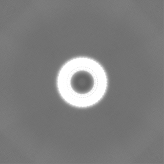
| Images |  emd_44895.png emd_44895.png | 135.9 KB | ||
| Filedesc metadata | emd-44895.cif.gz | 5.6 KB | ||
| Others | emd_44895_half_map_1.map.gzemd_44895_half_map_2.map.gz | 115.7 MB 115.7 MB | ||
| Archive directory |  http://ftp.pdbj.org/pub/emdb/structures/EMD-44895ftp://ftp.pdbj.org/pub/emdb/structures/EMD-44895 http://ftp.pdbj.org/pub/emdb/structures/EMD-44895ftp://ftp.pdbj.org/pub/emdb/structures/EMD-44895 | HTTPS FTP |
-Related structure data
| Related structure data |  9btqMC M: atomic model generated by this map C: citing same article ( |
|---|---|
| Similar structure data |
-Links
| EMDB pages | EMDB (EBI/PDBe) / EMDataResource |
|---|
-Map
| File | Download / File: emd_44895.map.gz / Format: CCP4 / Size: 125 MB / Type: IMAGE STORED AS FLOATING POINT NUMBER (4 BYTES) | ||||||||||||||||||||||||||||||||||||
|---|---|---|---|---|---|---|---|---|---|---|---|---|---|---|---|---|---|---|---|---|---|---|---|---|---|---|---|---|---|---|---|---|---|---|---|---|---|
| Annotation | cryo-EM map of extracellular tube from Microcystis Aeruginosa pcc 7806SL | ||||||||||||||||||||||||||||||||||||
| Projections & slices | Image control
Images are generated by Spider. | ||||||||||||||||||||||||||||||||||||
| Voxel size | X=Y=Z: 1.07 Å | ||||||||||||||||||||||||||||||||||||
| Density |
| ||||||||||||||||||||||||||||||||||||
| Symmetry | Space group: 1 | ||||||||||||||||||||||||||||||||||||
| Details | EMDB XML:
|
 Z (Sec.)
 Y (Row.)
 X (Col.)


-Supplemental data
-Half map: halfmap A
| File | emd_44895_half_map_1.map | ||||||||||||
|---|---|---|---|---|---|---|---|---|---|---|---|---|---|
| Annotation | halfmap A | ||||||||||||
| Projections & Slices |
| ||||||||||||
| Density Histograms |


-Half map: halfmap B
| File | emd_44895_half_map_2.map | ||||||||||||
|---|---|---|---|---|---|---|---|---|---|---|---|---|---|
| Annotation | halfmap B | ||||||||||||
| Projections & Slices |
| ||||||||||||
| Density Histograms |


- Sample components
-Entire : pili filaments
| Entire | Name: pili filaments |
|---|---|
| Components |
|
-Supramolecule #1: pili filaments
| Supramolecule | Name: pili filaments / type: complex / ID: 1 / Parent: 0 / Macromolecule list: all |
|---|---|
| Source (natural) | Organism: Microcystis aeruginosa PCC 7806SL (bacteria) |
-Macromolecule #1: pilin
| Macromolecule | Name: pilin / type: protein_or_peptide / ID: 1 / Number of copies: 1 / Enantiomer: LEVO |
|---|---|
| Source (natural) | Organism: Microcystis aeruginosa PCC 7806SL (bacteria) |
| Molecular weight | Theoretical: 15.515277 KDa |
| Sequence | String: MKASNYQEIA KAAILAGGLA AAGVLSVGES AQAQFIGTAS QSRVSAAVTS ILTDGNAAAT NSFAVEQVLP SSDYVFSGVV AVQVSYATT ISVGVGTAGA LTPVITAAEL TAPVVVNAGT QLTVERATAD AISKAATGSR FGDVSGIVRA WSAGTSVLD UniProtKB: Uncharacterized protein |
-Experimental details
-Structure determination
| Method | cryo EM |
|---|---|
 Processing Processing | helical reconstruction |
| Aggregation state | filament |
-Sample preparation
| Buffer | pH: 7.4 |
|---|---|
| Vitrification | Cryogen name: ETHANE |
- Electron microscopy
| Microscope | TFS KRIOS |
|---|---|
| Image recording | Film or detector model: GATAN K3 (6k x 4k) / Average electron dose: 50.0 e/Å2 |
| Electron beam | Acceleration voltage: 300 kV / Electron source:  FIELD EMISSION GUN FIELD EMISSION GUN |
| Electron optics | Illumination mode: FLOOD BEAM / Imaging mode: BRIGHT FIELD / Nominal defocus max: 2.0 µm / Nominal defocus min: 1.0 µm |
| Experimental equipment |  Model: Titan Krios / Image courtesy: FEI Company |
-Image processing
| Final reconstruction | Applied symmetry - Helical parameters - Δz: 3.69 Å Applied symmetry - Helical parameters - Δ&Phi: -14.74 ° Applied symmetry - Helical parameters - Axial symmetry: C1 (asymmetric) Resolution.type: BY AUTHOR / Resolution: 2.4 Å / Resolution method: FSC 0.143 CUT-OFF / Number images used: 1875465 |
|---|---|
| CTF correction | Type: PHASE FLIPPING AND AMPLITUDE CORRECTION |
| Startup model | Type of model: NONE |
| Final angle assignment | Type: NOT APPLICABLE |
