National Institutes of Health/National Center for Research Resources (NIH/NCRR)
United States
Citation
Journal: Traffic / Year: 2023 Title: GTP-stimulated membrane fission by the N-BAR protein AMPH-1. Authors: Lauren Kustigian / Xue Gong / Wei Gai / Jirapat Thongchol / Junjie Zhang / Jason Puchalla / Chavela M Carr / Hays S Rye / Abstract: Membrane-enclosed transport carriers sort biological molecules between stations in the cell in a dynamic process that is fundamental to the physiology of eukaryotic organisms. While much is known ...Membrane-enclosed transport carriers sort biological molecules between stations in the cell in a dynamic process that is fundamental to the physiology of eukaryotic organisms. While much is known about the formation and release of carriers from specific intracellular membranes, the mechanism of carrier formation from the recycling endosome, a compartment central to cellular signaling, remains to be resolved. In Caenorhabditis elegans, formation of transport carriers from the recycling endosome requires the dynamin-like, Eps15-homology domain (EHD) protein, RME-1, functioning with the Bin/Amphiphysin/Rvs (N-BAR) domain protein, AMPH-1. Here we show, using a free-solution single-particle technique known as burst analysis spectroscopy (BAS), that AMPH-1 alone creates small, tubular-vesicular products from large, unilamellar vesicles by membrane fission. Membrane fission requires the amphipathic H0 helix of AMPH-1 and is slowed in the presence of RME-1. Unexpectedly, AMPH-1-induced membrane fission is stimulated in the presence of GTP. Furthermore, the GTP-stimulated membrane fission activity seen for AMPH-1 is recapitulated by the heterodimeric N-BAR amphiphysin protein from yeast, Rvs161/167p, strongly suggesting that GTP-stimulated membrane fission is a general property of this important class of N-BAR proteins.
In the structure databanks used in Yorodumi, some data are registered as the other names, "COVID-19 virus" and "2019-nCoV". Here are the details of the virus and the list of structure data.
Jan 31, 2019. EMDB accession codes are about to change! (news from PDBe EMDB page)
EMDB accession codes are about to change! (news from PDBe EMDB page)
The allocation of 4 digits for EMDB accession codes will soon come to an end. Whilst these codes will remain in use, new EMDB accession codes will include an additional digit and will expand incrementally as the available range of codes is exhausted. The current 4-digit format prefixed with “EMD-” (i.e. EMD-XXXX) will advance to a 5-digit format (i.e. EMD-XXXXX), and so on. It is currently estimated that the 4-digit codes will be depleted around Spring 2019, at which point the 5-digit format will come into force.
The EM Navigator/Yorodumi systems omit the EMD- prefix.
Related info.:Q: What is EMD? / ID/Accession-code notation in Yorodumi/EM Navigator
Yorodumi is a browser for structure data from EMDB, PDB, SASBDB, etc.
This page is also the successor to EM Navigator detail page, and also detail information page/front-end page for Omokage search.
The word "yorodu" (or yorozu) is an old Japanese word meaning "ten thousand". "mi" (miru) is to see.
Related info.:EMDB / PDB / SASBDB / Comparison of 3 databanks / Yorodumi Search / Aug 31, 2016. New EM Navigator & Yorodumi / Yorodumi Papers / Jmol/JSmol / Function and homology information / Changes in new EM Navigator and Yorodumi
 Movie
Movie Controller


 Open data
 Basic information
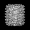
 Map data
 Sample
 Keywords
 Function and homology information
 Authors
 United States, 1 items
  Citation
 Structure visualization
 Downloads & links
 emd_44828.png
 http://ftp.pdbj.org/pub/emdb/structures/EMD-44828
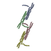

 Z (Sec.)
 Y (Row.)
 X (Col.)


 Sample components
 Processing
 Electron microscopy
 FIELD EMISSION GUN

