Movie
Movie Controller

[English] 日本語
 Yorodumi
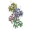
- EMDB-44777: CRYO-EM STRUCTURE OF CARDIAC MUSCLE ALPHA-ACTIN M305L HCM MUTANT -
+ Open data

- Basic information
| Entry | 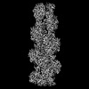 | |||||||||
|---|---|---|---|---|---|---|---|---|---|---|
| Title | CRYO-EM STRUCTURE OF CARDIAC MUSCLE ALPHA-ACTIN M305L HCM MUTANT | |||||||||
 Map data Map data | STRUCTURE OF CARDIAC MUSCLE ALPHA-ACTIN M305L HCM MUTANT | |||||||||
 Sample Sample |
| |||||||||
 Keywords Keywords | CYTOSKELETON / STRUCTURAL PROTEIN / filament | |||||||||
| Function / homology |  Function and homology information Function and homology informationcytoplasmic actin-based contraction involved in cell motility / actin-myosin filament sliding / cardiac myofibril assembly / actin filament-based movement / Formation of the dystrophin-glycoprotein complex (DGC) / cardiac muscle tissue morphogenesis / actomyosin structure organization / Striated Muscle Contraction / Regulation of CDH1 Function / I band ...cytoplasmic actin-based contraction involved in cell motility / actin-myosin filament sliding / cardiac myofibril assembly / actin filament-based movement / Formation of the dystrophin-glycoprotein complex (DGC) / cardiac muscle tissue morphogenesis / actomyosin structure organization / Striated Muscle Contraction / Regulation of CDH1 Function / I band / RHOB GTPase cycle / microfilament motor activity / myosin binding / heart contraction / mesenchyme migration / skeletal muscle thin filament assembly / RHOA GTPase cycle / cardiac muscle contraction / actin filament organization / sarcomere / filopodium / actin filament / Hydrolases; Acting on acid anhydrides; Acting on acid anhydrides to facilitate cellular and subcellular movement / lamellipodium / actin cytoskeleton / cell body / blood microparticle / response to ethanol / response to xenobiotic stimulus / focal adhesion / hydrolase activity / positive regulation of gene expression / negative regulation of apoptotic process / glutamatergic synapse / : / extracellular exosome / ATP binding / membrane / cytoplasm / cytosol Similarity search - Function | |||||||||
| Biological species | Mammalia (mammals) /  Homo sapiens (human) Homo sapiens (human) | |||||||||
| Method | single particle reconstruction / cryo EM / Resolution: 3.12 Å | |||||||||
 Authors Authors | Huang HL / Heissler SM / Chinthalapudi K | |||||||||
| Funding support |  United States, 1 items United States, 1 items
| |||||||||
 Citation Citation | Journal: To Be Published Title: CRYO-EM STRUCTURE OF CARDIAC MUSCLE ALPHA-ACTIN M305L HCM MUTANT Authors: Huang HL / Heissler SM / Chinthalapudi K | |||||||||
| History |
|
- Structure visualization
| Supplemental images |
|---|

- Downloads & links
-EMDB archive
| Map data | emd_44777.map.gz | 13 MB | EMDB map data format | |
|---|---|---|---|---|
| Header (meta data) | emd-44777-v30.xmlemd-44777.xml | 17.9 KB 17.9 KB | Display Display | EMDB header |
| FSC (resolution estimation) | emd_44777_fsc.xml | 8.5 KB | Display | FSC data file |
| Images |  emd_44777.png emd_44777.png | 111 KB | ||
| Filedesc metadata | emd-44777.cif.gz | 6 KB | ||
| Others | emd_44777_half_map_1.map.gzemd_44777_half_map_2.map.gz | 59.5 MB 59.5 MB | ||
| Archive directory |  http://ftp.pdbj.org/pub/emdb/structures/EMD-44777ftp://ftp.pdbj.org/pub/emdb/structures/EMD-44777 http://ftp.pdbj.org/pub/emdb/structures/EMD-44777ftp://ftp.pdbj.org/pub/emdb/structures/EMD-44777 | HTTPS FTP |
-Related structure data
| Related structure data |  9bphMC M: atomic model generated by this map C: citing same article ( |
|---|---|
| Similar structure data |
-Links
| EMDB pages | EMDB (EBI/PDBe) / EMDataResource |
|---|---|
| Related items in Molecule of the Month |


-Map
| File | Download / File: emd_44777.map.gz / Format: CCP4 / Size: 64 MB / Type: IMAGE STORED AS FLOATING POINT NUMBER (4 BYTES) | ||||||||||||||||||||||||||||||||||||
|---|---|---|---|---|---|---|---|---|---|---|---|---|---|---|---|---|---|---|---|---|---|---|---|---|---|---|---|---|---|---|---|---|---|---|---|---|---|
| Annotation | STRUCTURE OF CARDIAC MUSCLE ALPHA-ACTIN M305L HCM MUTANT | ||||||||||||||||||||||||||||||||||||
| Projections & slices | Image control
Images are generated by Spider. | ||||||||||||||||||||||||||||||||||||
| Voxel size | X=Y=Z: 0.891 Å | ||||||||||||||||||||||||||||||||||||
| Density |
| ||||||||||||||||||||||||||||||||||||
| Symmetry | Space group: 1 | ||||||||||||||||||||||||||||||||||||
| Details | EMDB XML:
|
 Z (Sec.)
 Y (Row.)
 X (Col.)


-Supplemental data
-Half map: Half Map A
| File | emd_44777_half_map_1.map | ||||||||||||
|---|---|---|---|---|---|---|---|---|---|---|---|---|---|
| Annotation | Half Map A | ||||||||||||
| Projections & Slices |
| ||||||||||||
| Density Histograms |


-Half map: Half Map B
| File | emd_44777_half_map_2.map | ||||||||||||
|---|---|---|---|---|---|---|---|---|---|---|---|---|---|
| Annotation | Half Map B | ||||||||||||
| Projections & Slices |
| ||||||||||||
| Density Histograms |


- Sample components
-Entire : CARDIAC ALPHA ACTIN M305L MUTANT
| Entire | Name: CARDIAC ALPHA ACTIN M305L MUTANT |
|---|---|
| Components |
|
-Supramolecule #1: CARDIAC ALPHA ACTIN M305L MUTANT
| Supramolecule | Name: CARDIAC ALPHA ACTIN M305L MUTANT / type: organelle_or_cellular_component / ID: 1 / Parent: 0 / Macromolecule list: #1 |
|---|---|
| Source (natural) | Organism: Mammalia (mammals) |
| Molecular weight | Theoretical: 168.076 KDa |
-Macromolecule #1: Actin, alpha cardiac muscle 1
| Macromolecule | Name: Actin, alpha cardiac muscle 1 / type: protein_or_peptide / ID: 1 / Number of copies: 4 / Enantiomer: LEVO EC number: Hydrolases; Acting on acid anhydrides; Acting on acid anhydrides to facilitate cellular and subcellular movement |
|---|---|
| Source (natural) | Organism: Homo sapiens (human) |
| Molecular weight | Theoretical: 41.825531 KDa |
| Recombinant expression | Organism: Mammalia (mammals) |
| Sequence | String: DDEETTALVC DNGSGLVKAG FAGDDAPRAV FPSIVGRPRH QGVMVGMGQK DSYVGDEAQS KRGILTLKYP IE(HIC)GII TNW DDMEKIWHHT FYNELRVAPE EHPTLLTEAP LNPKANREKM TQIMFETFNV PAMYVAIQAV LSLYASGRTT GIVLDSG DG VTHNVPIYEG ...String: DDEETTALVC DNGSGLVKAG FAGDDAPRAV FPSIVGRPRH QGVMVGMGQK DSYVGDEAQS KRGILTLKYP IE(HIC)GII TNW DDMEKIWHHT FYNELRVAPE EHPTLLTEAP LNPKANREKM TQIMFETFNV PAMYVAIQAV LSLYASGRTT GIVLDSG DG VTHNVPIYEG YALPHAIMRL DLAGRDLTDY LMKILTERGY SFVTTAEREI VRDIKEKLCY VALDFENEMA TAASSSSL E KSYELPDGQV ITIGNERFRC PETLFQPSFI GMESAGIHET TYNSIMKCDI DIRKDLYANN VLSGGTTLYP GIADRMQKE ITALAPSTMK IKIIAPPERK YSVWIGGSIL ASLSTFQQMW ISKQEYDEAG PSIVHRKCF UniProtKB: Actin, alpha cardiac muscle 1 |
-Macromolecule #2: MAGNESIUM ION
| Macromolecule | Name: MAGNESIUM ION / type: ligand / ID: 2 / Number of copies: 4 / Formula: MG |
|---|---|
| Molecular weight | Theoretical: 24.305 Da |
-Macromolecule #3: ADENOSINE-5'-DIPHOSPHATE
| Macromolecule | Name: ADENOSINE-5'-DIPHOSPHATE / type: ligand / ID: 3 / Number of copies: 4 / Formula: ADP |
|---|---|
| Molecular weight | Theoretical: 427.201 Da |
| Chemical component information |  ChemComp-ADP: |
-Experimental details
-Structure determination
| Method | cryo EM |
|---|---|
 Processing Processing | single particle reconstruction |
| Aggregation state | filament |
-Sample preparation
| Buffer | pH: 7 |
|---|---|
| Grid | Model: C-flat / Material: GOLD / Pretreatment - Type: GLOW DISCHARGE |
| Vitrification | Cryogen name: ETHANE |
- Electron microscopy
| Microscope | FEI TITAN KRIOS |
|---|---|
| Image recording | Film or detector model: GATAN K3 BIOQUANTUM (6k x 4k) / Number real images: 5275 / Average electron dose: 60.0 e/Å2 |
| Electron beam | Acceleration voltage: 300 kV / Electron source:  FIELD EMISSION GUN FIELD EMISSION GUN |
| Electron optics | Illumination mode: OTHER / Imaging mode: BRIGHT FIELD / Nominal defocus max: 2.5 µm / Nominal defocus min: 0.5 µm / Nominal magnification: 81000 |
| Sample stage | Specimen holder model: FEI TITAN KRIOS AUTOGRID HOLDER / Cooling holder cryogen: NITROGEN |
| Experimental equipment |  Model: Titan Krios / Image courtesy: FEI Company |