National Institutes of Health/National Institute of General Medical Sciences (NIH/NIGMS)
GM138863
United States
National Institutes of Health/National Institute of General Medical Sciences (NIH/NIGMS)
GM139876
United States
National Institutes of Health/National Institute of General Medical Sciences (NIH/NIGMS)
GM1454316
United States
Citation
Journal: J Am Chem Soc / Year: 2025 Title: Molecular Basis for the Activation of MsbA by Divalent Metals. Authors: Jixing Lyu / Hanieh Bahramimoghaddam / Tianqi Zhang / Elena Scott / Sangho D Yun / Gaya P Yadav / Minglei Zhao / David Russell / Arthur Laganowsky / Abstract: Proteins involved in the biogenesis of lipopolysaccharide (LPS), a lipid exclusive to Gram-negative bacteria, are promising candidates for drug discovery. Specifically, the ABC transporter MsbA plays ...Proteins involved in the biogenesis of lipopolysaccharide (LPS), a lipid exclusive to Gram-negative bacteria, are promising candidates for drug discovery. Specifically, the ABC transporter MsbA plays a crucial role in translocating an LPS precursor from the cytoplasmic to the periplasmic facing leaflet of the inner membrane, and small molecules that inhibit its function exhibit bactericidal activity. Here, we use native mass spectrometry (MS) to determine lipid binding affinities of MsbA from (PaMsbA), a Gram-negative bacteria associated with hospital-acquired infections, in different conformations. Unlike the transporter from , we show that the ATPase activity of PaMsbA is stimulated by Zn, Ni, and Mn and successfully trapping the protein with vanadate requires one of these metal ions. We also present cryogenic-electron microscopy structures of PaMsbA in occluded and open outward-facing conformations determined to resolutions of 2.58 and 2.44 Å, respectively. The structures reveal a triad of histidine residues, and mutation of these residues abolishes Zn binding and stimulation of PaMsbA activity by metal ions. Together, our studies provide insight into the structure of PaMsbA and its lipid binding preferences and reveal that a subset of divalent metals stimulates its ATPase activity.
In the structure databanks used in Yorodumi, some data are registered as the other names, "COVID-19 virus" and "2019-nCoV". Here are the details of the virus and the list of structure data.
Jan 31, 2019. EMDB accession codes are about to change! (news from PDBe EMDB page)
EMDB accession codes are about to change! (news from PDBe EMDB page)
The allocation of 4 digits for EMDB accession codes will soon come to an end. Whilst these codes will remain in use, new EMDB accession codes will include an additional digit and will expand incrementally as the available range of codes is exhausted. The current 4-digit format prefixed with “EMD-” (i.e. EMD-XXXX) will advance to a 5-digit format (i.e. EMD-XXXXX), and so on. It is currently estimated that the 4-digit codes will be depleted around Spring 2019, at which point the 5-digit format will come into force.
The EM Navigator/Yorodumi systems omit the EMD- prefix.
Related info.:Q: What is EMD? / ID/Accession-code notation in Yorodumi/EM Navigator
Yorodumi is a browser for structure data from EMDB, PDB, SASBDB, etc.
This page is also the successor to EM Navigator detail page, and also detail information page/front-end page for Omokage search.
The word "yorodu" (or yorozu) is an old Japanese word meaning "ten thousand". "mi" (miru) is to see.
Related info.:EMDB / PDB / SASBDB / Comparison of 3 databanks / Yorodumi Search / Aug 31, 2016. New EM Navigator & Yorodumi / Yorodumi Papers / Jmol/JSmol / Function and homology information / Changes in new EM Navigator and Yorodumi
 Movie
Movie Controller


 Open data
 Basic information
 Map data
 Sample
 Keywords
 Function and homology information
 Pseudomonas aeruginosa (bacteria)
 Authors
 United States, 3 items
  Citation
 Structure visualization
 Downloads & links
 emd_44444.png
 http://ftp.pdbj.org/pub/emdb/structures/EMD-44444


 Z (Sec.)
 Y (Row.)
 X (Col.)


 Sample components
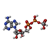
 Processing
 Electron microscopy
 FIELD EMISSION GUN


