 Movie
Movie Controller


+ Open data

- Basic information
| Entry | 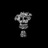 | |||||||||
|---|---|---|---|---|---|---|---|---|---|---|
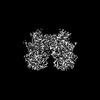
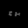
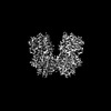
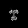
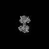
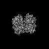
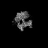
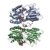
| Title | Full-length GC-A bound to ANP | |||||||||
 Map data Map data | ||||||||||
 Sample Sample |
| |||||||||
 Keywords Keywords | Single pass transmembrane protein / guanylyl cyclase / atrial natriuretic peptide receptor / hypertension / MEMBRANE PROTEIN | |||||||||
| Biological species |  Homo sapiens (human) Homo sapiens (human) | |||||||||
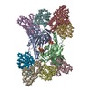
| Method | single particle reconstruction / cryo EM / Resolution: 7.4 Å | |||||||||
 Authors Authors | Liu S / Huang X | |||||||||
| Funding support |  United States, 1 items United States, 1 items
| |||||||||
 Citation Citation | Journal: Nat Struct Mol Biol / Year: 2025 Title: Architecture and activation of single-pass transmembrane receptor guanylyl cyclase. Authors: Shian Liu / Alexander M Payne / Jinan Wang / Lan Zhu / Navid Paknejad / Edward T Eng / Wei Liu / Yinglong Miao / Richard K Hite / Xin-Yun Huang / Abstract: The heart, in addition to its primary role in blood circulation, functions as an endocrine organ by producing cardiac hormone natriuretic peptides. These hormones regulate blood pressure through the ...The heart, in addition to its primary role in blood circulation, functions as an endocrine organ by producing cardiac hormone natriuretic peptides. These hormones regulate blood pressure through the single-pass transmembrane receptor guanylyl cyclase A (GC-A), also known as natriuretic peptide receptor 1. The binding of the peptide hormones to the extracellular domain of the receptor activates the intracellular guanylyl cyclase domain of the receptor to produce the second messenger cyclic guanosine monophosphate. Despite their importance, the detailed architecture and domain interactions within full-length GC-A remain elusive. Here we present cryo-electron microscopy structures, functional analyses and molecular dynamics simulations of full-length human GC-A, in both the absence and the presence of atrial natriuretic peptide. The data reveal the architecture of full-length GC-A, highlighting the spatial arrangement of its various functional domains. This insight is crucial for understanding how different parts of the receptor interact and coordinate during activation. The study elucidates the molecular basis of how extracellular signals are transduced across the membrane to activate the intracellular guanylyl cyclase domain. | |||||||||
| History |
|
- Structure visualization
| Supplemental images |
|---|

- Downloads & links
-EMDB archive
| Map data | emd_44437.map.gz | 25.5 MB |  EMDB map data format EMDB map data format | |
|---|---|---|---|---|
| Header (meta data) | emd-44437-v30.xmlemd-44437.xml | 13.5 KB 13.5 KB | Display Display | EMDB header |
| Images |  emd_44437.png emd_44437.png | 37.5 KB | ||
| Filedesc metadata | emd-44437.cif.gz | 5.2 KB | ||
| Others | emd_44437_half_map_1.map.gzemd_44437_half_map_2.map.gz | 25.1 MB 25.1 MB | ||
| Archive directory |  http://ftp.pdbj.org/pub/emdb/structures/EMD-44437ftp://ftp.pdbj.org/pub/emdb/structures/EMD-44437 http://ftp.pdbj.org/pub/emdb/structures/EMD-44437ftp://ftp.pdbj.org/pub/emdb/structures/EMD-44437 | HTTPS FTP |
-Related structure data


-Links
| EMDB pages | EMDB (EBI/PDBe) / EMDataResource |
|---|
-Map
| File | Download / File: emd_44437.map.gz / Format: CCP4 / Size: 27 MB / Type: IMAGE STORED AS FLOATING POINT NUMBER (4 BYTES) | ||||||||||||||||||||||||||||||||||||
|---|---|---|---|---|---|---|---|---|---|---|---|---|---|---|---|---|---|---|---|---|---|---|---|---|---|---|---|---|---|---|---|---|---|---|---|---|---|
| Projections & slices | Image control
Images are generated by Spider. | ||||||||||||||||||||||||||||||||||||
| Voxel size | X=Y=Z: 2.38 Å | ||||||||||||||||||||||||||||||||||||
| Density |
| ||||||||||||||||||||||||||||||||||||
| Symmetry | Space group: 1 | ||||||||||||||||||||||||||||||||||||
| Details | EMDB XML:
|
 Z (Sec.)
 Y (Row.)
 X (Col.)


-Supplemental data
-Half map: #1
| File | emd_44437_half_map_1.map | ||||||||||||
|---|---|---|---|---|---|---|---|---|---|---|---|---|---|
| Projections & Slices |
| ||||||||||||
| Density Histograms |


-Half map: #2
| File | emd_44437_half_map_2.map | ||||||||||||
|---|---|---|---|---|---|---|---|---|---|---|---|---|---|
| Projections & Slices |
| ||||||||||||
| Density Histograms |


- Sample components
-Entire : Atrial natriuretic peptide receptor 1 dimer
| Entire | Name: Atrial natriuretic peptide receptor 1 dimer |
|---|---|
| Components |
|
-Supramolecule #1: Atrial natriuretic peptide receptor 1 dimer
| Supramolecule | Name: Atrial natriuretic peptide receptor 1 dimer / type: complex / ID: 1 / Parent: 0 / Macromolecule list: all |
|---|---|
| Source (natural) | Organism: Homo sapiens (human) |
-Macromolecule #1: Atrial natriuretic peptide receptor 1
| Macromolecule | Name: Atrial natriuretic peptide receptor 1 / type: protein_or_peptide / ID: 1 / Enantiomer: LEVO |
|---|---|
| Source (natural) | Organism: Homo sapiens (human) |
| Sequence | String: DYKDDDDKAA A GNLTVAVV LPLANTSYPW SWARVGPAVE LALAQVKAR PDLLPGWTVR TVLGSSENAL GVCSDTAAPL AAVDLKWEHN PAVFLGPGCV Y AAAPVGRF TAHWRVPLLT AGAPALGFGV KDEYALTTRA GPSYAKLGDF VAALHRRLGW ER QALMLYA ...String: DYKDDDDKAA A GNLTVAVV LPLANTSYPW SWARVGPAVE LALAQVKAR PDLLPGWTVR TVLGSSENAL GVCSDTAAPL AAVDLKWEHN PAVFLGPGCV Y AAAPVGRF TAHWRVPLLT AGAPALGFGV KDEYALTTRA GPSYAKLGDF VAALHRRLGW ER QALMLYA YRPGDEEHCF FLVEGLFMRV RDRLNITVDH LEFAEDDLSH YTRLLRTMPR KGR VIYICS SPDAFRTLML LALEAGLCGE DYVFFHLDIF GQSLQGGQGP APRRPWERGD GQDV SARQA FQAAKIITYK DPDNPEYLEF LKQLKHLAYE QFNFTMEDGL VNTIPASFHD GLLLY IQAV TETLAHGGTV TDGENITQRM WNRSFQGVTG YLKIDSSGDR ETDFSLWDMD PENGAF RVV LNYNGTSQEL VAVSGRKLNW PLGYPPPDIP KCGFDNEDPA CNQDHLSTLE VLALVGS LS LLGILIVSFF IYRKMQLEKE LASELWRVRW EDVEPSSLER HLRSAGSRLT LSGRGSNY G SLLTTEGQFQ VFAKTAYYKG NLVAVKRVNR KRIELTRKVL FELKHMRDVQ NEHLTRFVG ACTDPPNICI LTEYCPRGSL QDILENESIT LDWMFRYSLT NDIVKGMLFL HNGAICSHGN LKSSNCVVD GRFVLKITDY GLESFRDLDP EQGHTVYAKK LWTAPELLRM ASPPVRGSQA G DVYSFGII LQEIALRSGV FHVEGLDLSP KEIIERVTRG EQPPFRPSLA LQSHLEELGL LM QRCWAED PQERPPFQQI RLTLRKFNRE NSSNILDNLL SRMEQYANNL EELVEERTQA YLE EKRKAE ALLYQILPHS VAEQLKRGET VQAEAFDSVT IYFSDIVGFT ALSAESTPMQ VVTL LNDLY TCFDAVIDNF DVYKVETIGD AYMVVSGLPV RNGRLHACEV ARMALALLDA VRSFR IRHR PQEQLRLRIG IHTGPVCAGV VGLKMPRYCL FGDTVNTASR MESNGEALKI HLSSET KAV LEEFGGFELE LRGDVEMKGK GKVRTYWLLG ERGSSTRG |
-Experimental details
-Structure determination
| Method | cryo EM |
|---|---|
 Processing Processing | single particle reconstruction |
| Aggregation state | particle |
-Sample preparation
| Concentration | 1 mg/mL |
|---|---|
| Buffer | pH: 8 |
| Vitrification | Cryogen name: ETHANE |
- Electron microscopy
| Microscope | TFS GLACIOS |
|---|---|
| Image recording | Film or detector model: FEI FALCON IV (4k x 4k) / Average electron dose: 40.0 e/Å2 |
| Electron beam | Acceleration voltage: 200 kV / Electron source:  FIELD EMISSION GUN FIELD EMISSION GUN |
| Electron optics | Illumination mode: FLOOD BEAM / Imaging mode: BRIGHT FIELD / Nominal defocus max: 1.5 µm / Nominal defocus min: 0.8 µm |
-Image processing
| Startup model | Type of model: NONE |
|---|---|
| Final reconstruction | Resolution.type: BY AUTHOR / Resolution: 7.4 Å / Resolution method: FSC 0.143 CUT-OFF / Number images used: 12270 |
| Initial angle assignment | Type: MAXIMUM LIKELIHOOD |
| Final angle assignment | Type: MAXIMUM LIKELIHOOD |
