 Movie
Movie Controller


+ Open data

- Basic information
| Entry | 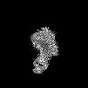 | |||||||||
|---|---|---|---|---|---|---|---|---|---|---|
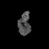
| Title | The map of the bent AlphaIIbBeta3 in complex with R21D10 Fab | |||||||||
 Map data Map data | It is the main map. | |||||||||
 Sample Sample |
| |||||||||
 Keywords Keywords | Murine monoclonal antibody R21D10 stabilizes a semi-extended conformation of the platelet AlphaIIbBeta3 integrin receptor and serves as an allosteric inhibitor of ligand binding and platelet aggregation. It also is the first mAb reported to inhibit the binding of protein disulfide isomerase. / BLOOD CLOTTING | |||||||||
| Biological species |  Homo sapiens (human) / Homo sapiens (human) /  | |||||||||
| Method | single particle reconstruction / cryo EM / Resolution: 4.4 Å | |||||||||
 Authors Authors | Wang JL / Walz T / Coller B / Wang L / Li JH | |||||||||
| Funding support |  United States, 1 items United States, 1 items
| |||||||||
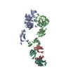
 Citation Citation | Journal: Blood Adv / Year: 2024 Title: An αIIbβ3 monoclonal antibody traps a semiextended conformation and allosterically inhibits large ligand binding. Authors: Lu Wang / Jialing Wang / Jihong Li / Thomas Walz / Barry S Coller / Abstract: Monoclonal antibodies (mAbs) have provided valuable information regarding the structure and function of platelet αIIbβ3. Protein disulfide isomerase (PDI) has been implicated in αIIbβ3 activation ...Monoclonal antibodies (mAbs) have provided valuable information regarding the structure and function of platelet αIIbβ3. Protein disulfide isomerase (PDI) has been implicated in αIIbβ3 activation and binds to thrombin-activated αIIbβ3. Using human platelets as the immunogen, we identified a new mAb (R21D10) that inhibits the binding of PDI to platelets activated with thrombin receptor-activating peptide (T6). R21D10 also partially inhibited T6-induced fibrinogen and PAC-1 binding to platelets, as well as T6- and adenosine 5'-diphosphate-induced platelet aggregation. Mutual competition experiments showed that R21D10 does not inhibit the binding of mAbs 10E5 (anti-αIIb cap domain) or 7E3 (anti-β3 β-I domain), and immunoblot studies indicated that R21D10 binds to β3. The dissociation of αIIbβ3 by EDTA had a minimal effect on R21D10 binding. Cryogenic electron microscopy of the αIIbβ3-R21D10 Fab complex revealed that R21D10 binds to the β3 integrin-epidermal growth factor 1 (I-EGF1) domain and traps an intermediate conformation of αIIbβ3 with semiextended leg domains. The binding of R21D10 produces a major structural change in the β3 I-EGF2 domain associated with a new interaction between the β3 I-EGF2 and αIIb thigh domains, which may prevent the swing-out motion of the β3 hybrid domain required for high-affinity ligand binding and protect αIIbβ3 from EDTA-induced dissociation. R21D10 partially reversed the ligand binding priming effect of eptifibatide, suggesting that it could convert the swung-out conformation into a semiextended conformation. We concluded that R21D10 inhibits ligand binding to αIIbβ3 via a unique allosteric mechanism, which may or may not be related to its inhibition of PDI binding. | |||||||||
| History |
|
- Structure visualization
| Supplemental images |
|---|

- Downloads & links
-EMDB archive
| Map data | emd_43983.map.gz | 230 MB |  EMDB map data format EMDB map data format | |
|---|---|---|---|---|
| Header (meta data) | emd-43983-v30.xmlemd-43983.xml | 22.1 KB 22.1 KB | Display Display | EMDB header |
| FSC (resolution estimation) | emd_43983_fsc.xml | 13.3 KB | Display | FSC data file |
| Images |  emd_43983.png emd_43983.png | 37.7 KB | ||
| Filedesc metadata | emd-43983.cif.gz | 6.3 KB | ||
| Others | emd_43983_half_map_1.map.gzemd_43983_half_map_2.map.gz | 226.8 MB 226.8 MB | ||
| Archive directory |  http://ftp.pdbj.org/pub/emdb/structures/EMD-43983ftp://ftp.pdbj.org/pub/emdb/structures/EMD-43983 http://ftp.pdbj.org/pub/emdb/structures/EMD-43983ftp://ftp.pdbj.org/pub/emdb/structures/EMD-43983 | HTTPS FTP |
-Related structure data
-Links
| EMDB pages | EMDB (EBI/PDBe) / EMDataResource |
|---|
-Map
| File | Download / File: emd_43983.map.gz / Format: CCP4 / Size: 244.1 MB / Type: IMAGE STORED AS FLOATING POINT NUMBER (4 BYTES) | ||||||||||||||||||||||||||||||||||||
|---|---|---|---|---|---|---|---|---|---|---|---|---|---|---|---|---|---|---|---|---|---|---|---|---|---|---|---|---|---|---|---|---|---|---|---|---|---|
| Annotation | It is the main map. | ||||||||||||||||||||||||||||||||||||
| Projections & slices | Image control
Images are generated by Spider. | ||||||||||||||||||||||||||||||||||||
| Voxel size | X=Y=Z: 0.86 Å | ||||||||||||||||||||||||||||||||||||
| Density |
| ||||||||||||||||||||||||||||||||||||
| Symmetry | Space group: 1 | ||||||||||||||||||||||||||||||||||||
| Details | EMDB XML:
|
 Z (Sec.)
 Y (Row.)
 X (Col.)


-Supplemental data
-Half map: It is the half B map.
| File | emd_43983_half_map_1.map | ||||||||||||
|---|---|---|---|---|---|---|---|---|---|---|---|---|---|
| Annotation | It is the half B map. | ||||||||||||
| Projections & Slices |
| ||||||||||||
| Density Histograms |


-Half map: It is the half A map.
| File | emd_43983_half_map_2.map | ||||||||||||
|---|---|---|---|---|---|---|---|---|---|---|---|---|---|
| Annotation | It is the half A map. | ||||||||||||
| Projections & Slices |
| ||||||||||||
| Density Histograms |


- Sample components
-Entire : The semi-extended AlphaIIbBeta3 in complex with R21D10 Fab
| Entire | Name: The semi-extended AlphaIIbBeta3 in complex with R21D10 Fab |
|---|---|
| Components |
|
-Supramolecule #1: The semi-extended AlphaIIbBeta3 in complex with R21D10 Fab
| Supramolecule | Name: The semi-extended AlphaIIbBeta3 in complex with R21D10 Fab type: complex / ID: 1 / Parent: 0 / Macromolecule list: all |
|---|---|
| Molecular weight | Theoretical: 250 KDa |
-Supramolecule #2: AlphaIIbBeta3 Integrin
| Supramolecule | Name: AlphaIIbBeta3 Integrin / type: complex / ID: 2 / Parent: 1 / Macromolecule list: #1-#2 |
|---|---|
| Source (natural) | Organism: Homo sapiens (human) |
-Supramolecule #3: R21D10 Fab
| Supramolecule | Name: R21D10 Fab / type: complex / ID: 3 / Parent: 1 / Macromolecule list: #3-#4 |
|---|---|
| Source (natural) | Organism: |
-Macromolecule #1: Integrin AlphaIIb
| Macromolecule | Name: Integrin AlphaIIb / type: protein_or_peptide / ID: 1 Details: The full-length Integrin AlphaIIb includes a signal peptide. Enantiomer: LEVO |
|---|---|
| Source (natural) | Organism: Homo sapiens (human) |
| Sequence | String: MARALCPLQA LWLLEWVLLL LGPCAAPPAW ALNLDPVQLT FYAGPNGSQF GFSLDFHKDS HGRVAIVVGA PRTLGPSQEE TGGVFLCPW RAEGGQCPSL LFDLRDETRN VGSQTLQTFK ARQGLGASVV SWSDVIVACA PWQHWNVLEK TEEAEKTPVG S CFLAQPES ...String: MARALCPLQA LWLLEWVLLL LGPCAAPPAW ALNLDPVQLT FYAGPNGSQF GFSLDFHKDS HGRVAIVVGA PRTLGPSQEE TGGVFLCPW RAEGGQCPSL LFDLRDETRN VGSQTLQTFK ARQGLGASVV SWSDVIVACA PWQHWNVLEK TEEAEKTPVG S CFLAQPES GRRAEYSPCR GNTLSRIYVE NDFSWDKRYC EAGFSSVVTQ AGELVLGAPG GYYFLGLLAQ APVADIFSSY RP GILLWHV SSQSLSFDSS NPEYFDGYWG YSVAVGEFDG DLNTTEYVVG APTWSWTLGA VEILDSYYQR LHRLRGEQMA SYF GHSVAV TDVNGDGRHD LLVGAPLYME SRADRKLAEV GRVYLFLQPR GPHALGAPSL LLTGTQLYGR FGSAIAPLGD LDRD GYNDI AVAAPYGGPS GRGQVLVFLG QSEGLRSRPS QVLDSPFPTG SAFGFSLRGA VDIDDNGYPD LIVGAYGANQ VAVYR AQPV VKASVQLLVQ DSLNPAVKSC VLPQTKTPVS CFNIQMCVGA TGHNIPQKLS LNAELQLDRQ KPRQGRRVLL LGSQQA GTT LNLDLGGKHS PICHTTMAFL RDEADFRDKL SPIVLSLNVS LPPTEAGMAP AVVLHGDTHV QEQTRIVLDC GEDDVCV PQ LQLTASVTGS PLLVGADNVL ELQMDAANEG EGAYEAELAV HLPQGAHYMR ALSNVEGFER LICNQKKENE TRVVLCEL G NPMKKNAQIG IAMLVSVGNL EEAGESVSFQ LQIRSKNSQN PNSKIVLLDV PVRAEAQVEL RGNSFPASLV VAAEEGERE QNSLDSWGPK VEHTYELHNN GPGTVNGLHL SIHLPGQSQP SDLLYILDIQ PQGGLQCFPQ PPVNPLKVDW GLPIPSPSPI HPAHHKRDR RQIFLPEPEQ PSRLQDPVLV SCDSAPCTVV QCDLQEMARG QRAMVTVLAF LWLPSLYQRP LDQFVLQSHA W FNVSSLPY AVPPLSLPRG EAQVWTQLLR ALEERAIPIW WVLVGVLGGL LLLTILVLAM WKVGFFKRNR PPLEEDDEEG E |
-Macromolecule #2: Integrin Beta3
| Macromolecule | Name: Integrin Beta3 / type: protein_or_peptide / ID: 2 Details: The sequence is for the full-length Beta3 including a signal peptide. Enantiomer: LEVO |
|---|---|
| Source (natural) | Organism: Homo sapiens (human) |
| Sequence | String: MRARPRPRPL WATVLALGAL AGVGVGGPNI CTTRGVSSCQ QCLAVSPMCA WCSDEALPLG SPRCDLKENL LKDNCAPESI EFPVSEARV LEDRPLSDKG SGDSSQVTQV SPQRIALRLR PDDSKNFSIQ VRQVEDYPVD IYYLMDLSYS MKDDLWSIQN L |
-Macromolecule #3: R21D10 Fab heavy chain
| Macromolecule | Name: R21D10 Fab heavy chain / type: protein_or_peptide / ID: 3 / Enantiomer: LEVO |
|---|---|
| Source (natural) | Organism: |
| Sequence | String: EVQLQESGAE LAKPGASVKM SCKASGYTFT DYWMHWVKQR PGQGLEWIGY INPSTGYTEY NQKFKDKATL TADKSSSTAY MQLSSLTSE DSAVYYCARW RGVYRSDVHY YAMDYWGQGT SVTVSSAKTT APSVYPLAPV CGDTTGSSVT LGCLVKGYFP E |
-Macromolecule #4: R211010 Fab light chain
| Macromolecule | Name: R211010 Fab light chain / type: protein_or_peptide / ID: 4 / Enantiomer: LEVO |
|---|---|
| Source (natural) | Organism: |
| Sequence | String: QIVLTQSPAI MSASPGEKVT MTCSASSSVS YMHWYQQKSG TSPKRWIYDT SKLASGVPAR FSGSGSGTSY SLTISSMEAE DAATYYCQQ WSSKPPTFGA GTKLELKWAD AAPTVSIFPP SSEQLTSGGA SVVCFLNNFY PKDINVKWKI DGSERQNGVL N SWTDQDSK ...String: QIVLTQSPAI MSASPGEKVT MTCSASSSVS YMHWYQQKSG TSPKRWIYDT SKLASGVPAR FSGSGSGTSY SLTISSMEAE DAATYYCQQ WSSKPPTFGA GTKLELKWAD AAPTVSIFPP SSEQLTSGGA SVVCFLNNFY PKDINVKWKI DGSERQNGVL N SWTDQDSK DSTYSMSSTL TLTKDEYERH NSYTCEATHK TSTSPIVKSF NRNEC |
-Experimental details
-Structure determination
| Method | cryo EM |
|---|---|
 Processing Processing | single particle reconstruction |
| Aggregation state | particle |
-Sample preparation
| Concentration | 0.05 mg/mL |
|---|---|
| Buffer | pH: 7.4 |
| Grid | Model: Quantifoil R1.2/1.3 / Material: GRAPHENE OXIDE / Mesh: 400 / Support film - Material: GRAPHENE OXIDE / Support film - topology: CONTINUOUS / Pretreatment - Type: GLOW DISCHARGE / Pretreatment - Time: 5 sec. |
| Vitrification | Cryogen name: ETHANE / Chamber humidity: 100 % / Chamber temperature: 277 K / Instrument: FEI VITROBOT MARK I |
| Details | AlphaIIbBeta3 in complex with R21D10 Fab |
- Electron microscopy
| Microscope | FEI TITAN KRIOS |
|---|---|
| Image recording | Film or detector model: GATAN K3 (6k x 4k) / Average electron dose: 54.0 e/Å2 |
| Electron beam | Acceleration voltage: 300 kV / Electron source:  FIELD EMISSION GUN FIELD EMISSION GUN |
| Electron optics | Illumination mode: FLOOD BEAM / Imaging mode: BRIGHT FIELD / Nominal defocus max: 2.5 µm / Nominal defocus min: 1.5 µm / Nominal magnification: 81000 |
| Sample stage | Specimen holder model: FEI TITAN KRIOS AUTOGRID HOLDER / Cooling holder cryogen: NITROGEN |
| Experimental equipment |  Model: Titan Krios / Image courtesy: FEI Company |
+Image processing

-Atomic model buiding 1
| Initial model |
| ||||||||||
|---|---|---|---|---|---|---|---|---|---|---|---|
| Refinement | Space: REAL / Protocol: RIGID BODY FIT |

