 ムービー
ムービー コントローラー


+ データを開く

- 基本情報
基本情報
| 登録情報 |  | |||||||||
|---|---|---|---|---|---|---|---|---|---|---|
| タイトル | Structure of the auto-fluorescent membrane-bound red body organelle from Nannochloropsis oceanica in situ | |||||||||
 マップデータ マップデータ | Tomographic reconstruction of a cryo-lamella through a Nannochloropsis oceanica cell showing a red body within its cellular context. | |||||||||
 試料 試料 |
| |||||||||
 キーワード キーワード | Organelle / Algae / Intracellular Transport / Carotenoid / LIPID TRANSPORT | |||||||||
| 生物種 |  Nannochloropsis oceanica (真核生物) Nannochloropsis oceanica (真核生物) | |||||||||
| 手法 | 電子線トモグラフィー法 / クライオ電子顕微鏡法 | |||||||||
 データ登録者 データ登録者 | Grob P / Danielle J / Gee CW | |||||||||
| 資金援助 |  米国, 2件 米国, 2件
| |||||||||
 引用 引用 | ジャーナル: Nat Commun / 年: 2024 タイトル: Implicating the red body of Nannochloropsis in forming the recalcitrant cell wall polymer algaenan. 著者: Christopher W Gee / Johan Andersen-Ranberg / Ethan Boynton / Rachel Z Rosen / Danielle Jorgens / Patricia Grob / Hoi-Ying N Holman / Krishna K Niyogi /  要旨: Stramenopile algae contribute significantly to global primary productivity, and one class, Eustigmatophyceae, is increasingly studied for applications in high-value lipid production. Yet much about ...Stramenopile algae contribute significantly to global primary productivity, and one class, Eustigmatophyceae, is increasingly studied for applications in high-value lipid production. Yet much about their basic biology remains unknown, including the nature of an enigmatic, pigmented globule found in vegetative cells. Here, we present an in-depth examination of this "red body," focusing on Nannochloropsis oceanica. During the cell cycle, the red body forms adjacent to the plastid, but unexpectedly it is secreted and released with the autosporangial wall following cell division. Shed red bodies contain antioxidant ketocarotenoids, and overexpression of a beta-carotene ketolase results in enlarged red bodies. Infrared spectroscopy indicates long-chain, aliphatic lipids in shed red bodies and cell walls, and UHPLC-HRMS detects a C32 alkyl diol, a potential precursor of algaenan, a recalcitrant cell wall polymer. We propose that the red body transports algaenan precursors from plastid to apoplast to be incorporated into daughter cell walls. | |||||||||
| 履歴 |
|
- 構造の表示
構造の表示
| 添付画像 |
|---|

- ダウンロードとリンク
-EMDBアーカイブ
| マップデータ | emd_43893.map.gz | 514.9 MB |  EMDBマップデータ形式 EMDBマップデータ形式 | |
|---|---|---|---|---|
| ヘッダ (付随情報) | emd-43893-v30.xmlemd-43893.xml | 12.7 KB 12.7 KB | 表示 表示 | EMDBヘッダ |
| 画像 | 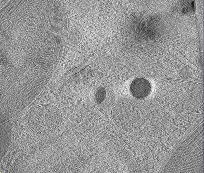 emd_43893.png emd_43893.png | 199.2 KB | ||
| Filedesc metadata | emd-43893.cif.gz | 5 KB | ||
| アーカイブディレクトリ |  http://ftp.pdbj.org/pub/emdb/structures/EMD-43893ftp://ftp.pdbj.org/pub/emdb/structures/EMD-43893 http://ftp.pdbj.org/pub/emdb/structures/EMD-43893ftp://ftp.pdbj.org/pub/emdb/structures/EMD-43893 | HTTPS FTP |
-検証レポート
| 文書・要旨 | emd_43893_validation.pdf.gz | 497.1 KB | 表示 | EMDB検証レポート |
|---|---|---|---|---|
| 文書・詳細版 | emd_43893_full_validation.pdf.gz | 496.6 KB | 表示 | |
| XML形式データ | emd_43893_validation.xml.gz | 4.5 KB | 表示 | |
| CIF形式データ | emd_43893_validation.cif.gz | 4.9 KB | 表示 | |
| アーカイブディレクトリ | https://ftp.pdbj.org/pub/emdb/validation_reports/EMD-43893ftp://ftp.pdbj.org/pub/emdb/validation_reports/EMD-43893 | HTTPS FTP |
-リンク
| EMDBのページ | EMDB (EBI/PDBe) / EMDataResource |
|---|
-マップ
| ファイル | ダウンロード / ファイル: emd_43893.map.gz / 形式: CCP4 / 大きさ: 558.8 MB / タイプ: IMAGE STORED AS FLOATING POINT NUMBER (4 BYTES) | ||||||||||||||||||||||||||||||||
|---|---|---|---|---|---|---|---|---|---|---|---|---|---|---|---|---|---|---|---|---|---|---|---|---|---|---|---|---|---|---|---|---|---|
| 注釈 | Tomographic reconstruction of a cryo-lamella through a Nannochloropsis oceanica cell showing a red body within its cellular context. | ||||||||||||||||||||||||||||||||
| 投影像・断面図 | 画像のコントロール
画像は Spider により作成 これらの図は立方格子座標系で作成されたものです | ||||||||||||||||||||||||||||||||
| ボクセルのサイズ | X=Y=Z: 22.13 Å | ||||||||||||||||||||||||||||||||
| 密度 |
| ||||||||||||||||||||||||||||||||
| 対称性 | 空間群: 1 | ||||||||||||||||||||||||||||||||
| 詳細 | EMDB XML:
|
 Z (Sec.)
 Y (Row.)
 X (Col.)


-添付データ
- 試料の構成要素
-全体 : Nannochloropsis oceanica CCMP1779
| 全体 | 名称: Nannochloropsis oceanica CCMP1779 |
|---|---|
| 要素 |
|
-超分子 #1: Nannochloropsis oceanica CCMP1779
| 超分子 | 名称: Nannochloropsis oceanica CCMP1779 / タイプ: cell / ID: 1 / 親要素: 0 詳細: Tomographic reconstruction of a red body organelle from Nannochloropsis oceanica in situ |
|---|---|
| 由来(天然) | 生物種: Nannochloropsis oceanica (真核生物) / 株: CCMP1779 |
-実験情報
-構造解析
| 手法 | クライオ電子顕微鏡法 |
|---|---|
 解析 解析 | 電子線トモグラフィー法 |
| 試料の集合状態 | cell |
-試料調製
| 緩衝液 | pH: 8.1 構成要素:
| ||||||||||||
|---|---|---|---|---|---|---|---|---|---|---|---|---|---|
| グリッド | モデル: Quantifoil R2/2 / 材質: GOLD / メッシュ: 300 / 支持フィルム - 材質: CARBON / 支持フィルム - トポロジー: HOLEY ARRAY / 支持フィルム - Film thickness: 12 / 前処理 - タイプ: PLASMA CLEANING / 前処理 - 時間: 60 sec. / 前処理 - 雰囲気: AIR / 前処理 - 気圧: 0.027 kPa / 詳細: the grid was soaked in chloroform before use | ||||||||||||
| 凍結 | 凍結剤: ETHANE / チャンバー内湿度: 100 % / チャンバー内温度: 283 K / 装置: FEI VITROBOT MARK IV / 詳細: Blotted manually from opposite side of the grid. | ||||||||||||
| 詳細 | Nannochloropsis oceanica CCMP1779 cells were grown in artificial seawater and f-media enrichment entrained to a 12-12 light-dark photoperiod and sampled in mid-log phase (~1x10^7 cells/ml), shortly after subjective dark when cells are undergoing division. | ||||||||||||
| 切片作成 | 集束イオンビーム - 装置: OTHER / 集束イオンビーム - イオン: OTHER / 集束イオンビーム - 電圧: 30 / 集束イオンビーム - 電流: 0.037 / 集束イオンビーム - 時間: 600 / 集束イオンビーム - 温度: 123 K / 集束イオンビーム - Initial thickness: 1000 / 集束イオンビーム - 最終 厚さ: 380 集束イオンビーム - 詳細: Grids were transfered to a Leica Ace 900 (Leica Microsystems) for coating with 5nm of platinum prior to being transferred to the Zeiss Crossbeam 540 (Zeiss, Germany) ...集束イオンビーム - 詳細: Grids were transfered to a Leica Ace 900 (Leica Microsystems) for coating with 5nm of platinum prior to being transferred to the Zeiss Crossbeam 540 (Zeiss, Germany). The Zeiss Crossbeam 540 operated with a Leica CryoStage (Leica Microsystems, GmbH) cooled to -150 deg. C was used for milling and imaging of the frozen cells. To create lamellae used for cryo-tomography, milling was performed with a gallium ion source at an energy of 37 pA and a working distance of 5mm. Imaging of the grid and milled lamellae was done using an Everhart-Thornley detector at 2.0 kV.. The value given for _em_focused_ion_beam.instrument is Zeiss Crossbeam 540. This is not in a list of allowed values {'OTHER', 'DB235'} so OTHER is written into the XML file. |
- 電子顕微鏡法
| 顕微鏡 | TFS KRIOS |
|---|---|
| 特殊光学系 | エネルギーフィルター - 名称: GIF Bioquantum / エネルギーフィルター - スリット幅: 35 eV |
| 撮影 | フィルム・検出器のモデル: GATAN K3 (6k x 4k) / 平均電子線量: 100.0 e/Å2 詳細: images were collected in movie mode and super resolution |
| 電子線 | 加速電圧: 300 kV / 電子線源:  FIELD EMISSION GUN FIELD EMISSION GUN |
| 電子光学系 | C2レンズ絞り径: 100.0 µm / 照射モード: FLOOD BEAM / 撮影モード: BRIGHT FIELD / Cs: 2.7 mm / 最大 デフォーカス(公称値): 6.0 µm / 最小 デフォーカス(公称値): 1.6 µm / 倍率(公称値): 15000 |
| 試料ステージ | 試料ホルダーモデル: FEI TITAN KRIOS AUTOGRID HOLDER ホルダー冷却材: NITROGEN |
| 実験機器 |  モデル: Titan Krios / 画像提供: FEI Company |
-画像解析
| 詳細 | the movie frames were aligned with MotionCor2 and binned to normal resolution |
|---|---|
| 最終 再構成 | アルゴリズム: BACK PROJECTION / ソフトウェア - 名称: IMOD (ver. 4.11.24) / ソフトウェア - 詳細: Etomo / 使用した粒子像数: 61 |
