 Movie
Movie Controller

[English] 日本語
 Yorodumi
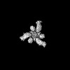
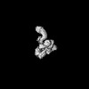
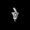
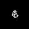
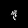
- EMDB-43886: Negative stain EM map of SARS-COV-2 (XBB) spike protein in comple... -
+ Open data

- Basic information
| Entry | 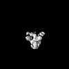 | |||||||||
|---|---|---|---|---|---|---|---|---|---|---|
| Title | Negative stain EM map of SARS-COV-2 (XBB) spike protein in complex with Fab COV2-3906 | |||||||||
 Map data Map data | ||||||||||
 Sample Sample |
| |||||||||
 Keywords Keywords | VIRAL-PROTEIN / Fab / IMMUNE-complex / VIRAL PROTEIN / VIRAL PROTEIN-IMMUNE SYSTEM complex | |||||||||
| Biological species |   Severe acute respiratory syndrome coronavirus 2 / Severe acute respiratory syndrome coronavirus 2 /  Homo sapiens (human) Homo sapiens (human) | |||||||||
| Method | single particle reconstruction / negative staining / Resolution: 25.0 Å | |||||||||
 Authors Authors | Binshtein E / Crowe JE | |||||||||
| Funding support |  United States, 1 items United States, 1 items
| |||||||||
 Citation Citation | Journal: Nat Microbiol / Year: 2026 Title: Epitope-focused discovery of SARS-CoV-2 antibodies that potently neutralize Omicron variants. Authors: Seth J Zost / Naveenchandra Suryadevara / Lauren E Williamson / Suzanne M Scheaffer / Elad Binshtein / Cameron D Buchman / Nicole V Johnson / Nicholas J Catanzaro / Silvia Ravera / Nathaniel ...Authors: Seth J Zost / Naveenchandra Suryadevara / Lauren E Williamson / Suzanne M Scheaffer / Elad Binshtein / Cameron D Buchman / Nicole V Johnson / Nicholas J Catanzaro / Silvia Ravera / Nathaniel S Chapman / Luke Myers / Ajit R Ramamohan / Laura S Handal / Doan C Nguyen / Andrew Trivette / James R Martinez / Eduardo Villalobos / Stacey A Rutherford / F Eun-Hyung Lee / Alexandra Schäfer / Ralph S Baric / Jason S McLellan / Michael S Diamond / Robert H Carnahan / James E Crowe / Abstract: The emergence of SARS-CoV-2 Omicron variants has led to viral escape from many clinically approved monoclonal antibodies (mAbs) due to rapid evolution of the receptor-binding domain (RBD). Co- ...The emergence of SARS-CoV-2 Omicron variants has led to viral escape from many clinically approved monoclonal antibodies (mAbs) due to rapid evolution of the receptor-binding domain (RBD). Co-circulation of SARS-CoV-2 variants with unique sets of antigenic substitutions has further complicated therapeutic mAb discovery. New approaches are needed to rapidly discover and characterize mAbs with preferred specificity and functional characteristics. Here we describe and perform epitope-focused mAb discovery using glycan-masked antigens. We isolated and expressed a panel of 303 mAbs, some of which potently neutralize divergent Omicron subvariants by targeting the class 3 antigenic site on SARS-CoV-2 RBD. Epitope mapping of these antibodies revealed a spectrum of cross-reactivity and differential recognition of the class 3 site, validating the utility of this enrichment approach for targeted mAb discovery. Together, this work rationally designs glycan-masked engineered RBDs and uses them to isolate mAbs that potently neutralize antigenically divergent SARS-CoV-2 variants. | |||||||||
| History |
|
- Structure visualization
| Supplemental images |
|---|

- Downloads & links
-EMDB archive
| Map data | emd_43886.map.gz | 7.5 MB |  EMDB map data format EMDB map data format | |
|---|---|---|---|---|
| Header (meta data) | emd-43886-v30.xmlemd-43886.xml | 17.5 KB 17.5 KB | Display Display | EMDB header |
| Images |  emd_43886.png emd_43886.png | 29.7 KB | ||
| Filedesc metadata | emd-43886.cif.gz | 4.8 KB | ||
| Others | emd_43886_half_map_1.map.gzemd_43886_half_map_2.map.gz | 7.4 MB 7.4 MB | ||
| Archive directory |  http://ftp.pdbj.org/pub/emdb/structures/EMD-43886ftp://ftp.pdbj.org/pub/emdb/structures/EMD-43886 http://ftp.pdbj.org/pub/emdb/structures/EMD-43886ftp://ftp.pdbj.org/pub/emdb/structures/EMD-43886 | HTTPS FTP |
-Related structure data


-Links
| EMDB pages | EMDB (EBI/PDBe) / EMDataResource |
|---|
-Map
| File | Download / File: emd_43886.map.gz / Format: CCP4 / Size: 8 MB / Type: IMAGE STORED AS FLOATING POINT NUMBER (4 BYTES) | ||||||||||||||||||||||||||||||||||||
|---|---|---|---|---|---|---|---|---|---|---|---|---|---|---|---|---|---|---|---|---|---|---|---|---|---|---|---|---|---|---|---|---|---|---|---|---|---|
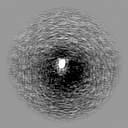
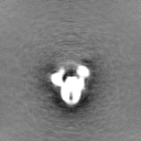
| Projections & slices | Image control
Images are generated by Spider. | ||||||||||||||||||||||||||||||||||||
| Voxel size | X=Y=Z: 4.36 Å | ||||||||||||||||||||||||||||||||||||
| Density |
| ||||||||||||||||||||||||||||||||||||
| Symmetry | Space group: 1 | ||||||||||||||||||||||||||||||||||||
| Details | EMDB XML:
|
 Z (Sec.)
 Y (Row.)
 X (Col.)


-Supplemental data
-Half map: #1
| File | emd_43886_half_map_1.map | ||||||||||||
|---|---|---|---|---|---|---|---|---|---|---|---|---|---|
| Projections & Slices |
| ||||||||||||
| Density Histograms |


-Half map: #2
| File | emd_43886_half_map_2.map | ||||||||||||
|---|---|---|---|---|---|---|---|---|---|---|---|---|---|
| Projections & Slices |
| ||||||||||||
| Density Histograms |


- Sample components
-Entire : SARS-COV-2 (XBB) spike in complex with Fab COV2-3872
| Entire | Name: SARS-COV-2 (XBB) spike in complex with Fab COV2-3872 |
|---|---|
| Components |
|
-Supramolecule #1: SARS-COV-2 (XBB) spike in complex with Fab COV2-3872
| Supramolecule | Name: SARS-COV-2 (XBB) spike in complex with Fab COV2-3872 / type: complex / ID: 1 / Parent: 0 |
|---|---|
| Molecular weight | Theoretical: 540 MDa |
-Supramolecule #2: Cov2-spike
| Supramolecule | Name: Cov2-spike / type: complex / ID: 2 / Parent: 1 |
|---|---|
| Source (natural) | Organism: Severe acute respiratory syndrome coronavirus 2 |
-Supramolecule #3: Fab
| Supramolecule | Name: Fab / type: complex / ID: 3 / Parent: 1 |
|---|---|
| Source (natural) | Organism: Homo sapiens (human) |
| Molecular weight | Theoretical: 50 KDa |
-Experimental details
-Structure determination
| Method | negative staining |
|---|---|
 Processing Processing | single particle reconstruction |
| Aggregation state | particle |
-Sample preparation
| Concentration | 0.01 mg/mL |
|---|---|
| Buffer | pH: 8 |
| Staining | Type: NEGATIVE / Material: uranyl formate |
| Grid | Model: Quantifoil / Material: COPPER / Mesh: 400 / Support film - Material: CARBON / Support film - topology: CONTINUOUS |
- Electron microscopy
| Microscope | FEI TECNAI F20 |
|---|---|
| Image recording | Film or detector model: GATAN ULTRASCAN 4000 (4k x 4k) / Detector mode: COUNTING / Number grids imaged: 1 / Number real images: 146 / Average exposure time: 1.0 sec. / Average electron dose: 35.0 e/Å2 |
| Electron beam | Acceleration voltage: 200 kV / Electron source:  FIELD EMISSION GUN FIELD EMISSION GUN |
| Electron optics | C2 aperture diameter: 100.0 µm / Illumination mode: OTHER / Imaging mode: BRIGHT FIELD / Cs: 2.2 mm / Nominal defocus max: 1.6 µm / Nominal defocus min: 1.0 µm / Nominal magnification: 50000 |
| Sample stage | Specimen holder model: SIDE ENTRY, EUCENTRIC |
| Experimental equipment |  Model: Tecnai F20 / Image courtesy: FEI Company |
+Image processing
-Atomic model buiding 1
| Refinement | Space: REAL |
|---|
