 Movie
Movie Controller

[English] 日本語
 Yorodumi
- EMDB-43846: Structure of Circularly Permuted 50S Ribosomal Subunit Assembly I... -
+ Open data

- Basic information
| Entry |  | ||||||||||||
|---|---|---|---|---|---|---|---|---|---|---|---|---|---|
| Title | Structure of Circularly Permuted 50S Ribosomal Subunit Assembly Intermediate - CP45 Class C2 | ||||||||||||
 Map data Map data | Class C2 CP45 50S Ribosomal Intermediate | ||||||||||||
 Sample Sample |
| ||||||||||||
 Keywords Keywords | 50S subunit / assembly intermediate / RNA-protein complex / ribosome | ||||||||||||
| Biological species |  | ||||||||||||
| Method | single particle reconstruction / cryo EM / Resolution: 4.8 Å | ||||||||||||
 Authors Authors | Dong X / Sheng K / Williamson JR | ||||||||||||
| Funding support |  United States, 3 items United States, 3 items
| ||||||||||||
 Citation Citation | Journal: Nucleic Acids Res / Year: 2024 Title: Assembly of the bacterial ribosome with circularly permuted rRNA. Authors: Xiyu Dong / Kai Sheng / Luca F R Gebert / Sriram Aiyer / Ian J MacRae / Dmitry Lyumkis / James R Williamson / Abstract: Co-transcriptional assembly is an integral feature of the formation of RNA-protein complexes that mediate translation. For ribosome synthesis, prior studies have indicated that the strict order of ...Co-transcriptional assembly is an integral feature of the formation of RNA-protein complexes that mediate translation. For ribosome synthesis, prior studies have indicated that the strict order of transcription of rRNA domains may not be obligatory during bacterial ribosome biogenesis, since a series of circularly permuted rRNAs are viable. In this work, we report the structural insights into assembly of the bacterial ribosome large subunit (LSU) based on cryo-EM density maps of intermediates that accumulate during in vitro ribosome synthesis using a set of circularly permuted (CiPer) rRNAs. The observed ensemble of 23 resolved ribosome large subunit intermediates reveals conserved assembly routes with an underlying hierarchy among cooperative assembly blocks. There are intricate interdependencies for the formation of key structural rRNA helices revealed from the circular permutation of rRNA. While the order of domain synthesis is not obligatory, the order of domain association does appear to proceed with a particular order, likely due to the strong evolutionary pressure on efficient ribosome synthesis. This work reinforces the robustness of the known assembly hierarchy of the bacterial large ribosomal subunit and offers a coherent view of how efficient assembly of CiPer rRNAs can be understood in that context. | ||||||||||||
| History |
|
- Structure visualization
| Supplemental images |
|---|

- Downloads & links
-EMDB archive
| Map data | emd_43846.map.gz | 93.1 MB |  EMDB map data format EMDB map data format | |
|---|---|---|---|---|
| Header (meta data) | emd-43846-v30.xmlemd-43846.xml | 16.5 KB 16.5 KB | Display Display | EMDB header |
| FSC (resolution estimation) | emd_43846_fsc.xml | 12.2 KB | Display | FSC data file |
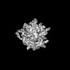
| Images | 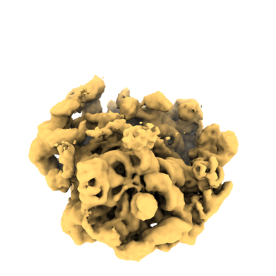 emd_43846.png emd_43846.png | 95.5 KB | ||
| Filedesc metadata | emd-43846.cif.gz | 4.6 KB | ||
| Others | emd_43846_half_map_1.map.gzemd_43846_half_map_2.map.gz | 173.8 MB 173.8 MB | ||
| Archive directory |  http://ftp.pdbj.org/pub/emdb/structures/EMD-43846ftp://ftp.pdbj.org/pub/emdb/structures/EMD-43846 http://ftp.pdbj.org/pub/emdb/structures/EMD-43846ftp://ftp.pdbj.org/pub/emdb/structures/EMD-43846 | HTTPS FTP |
-Related structure data
| Related structure data | C: citing same article ( |
|---|


-Links
| EMDB pages | EMDB (EBI/PDBe) / EMDataResource |
|---|
-Map
| File | Download / File: emd_43846.map.gz / Format: CCP4 / Size: 187 MB / Type: IMAGE STORED AS FLOATING POINT NUMBER (4 BYTES) | ||||||||||||||||||||||||||||||||||||
|---|---|---|---|---|---|---|---|---|---|---|---|---|---|---|---|---|---|---|---|---|---|---|---|---|---|---|---|---|---|---|---|---|---|---|---|---|---|
| Annotation | Class C2 CP45 50S Ribosomal Intermediate | ||||||||||||||||||||||||||||||||||||
| Projections & slices | Image control
Images are generated by Spider. | ||||||||||||||||||||||||||||||||||||
| Voxel size | X=Y=Z: 1.15 Å | ||||||||||||||||||||||||||||||||||||
| Density |
| ||||||||||||||||||||||||||||||||||||
| Symmetry | Space group: 1 | ||||||||||||||||||||||||||||||||||||
| Details | EMDB XML:
|
 Z (Sec.)
 Y (Row.)
 X (Col.)


-Supplemental data
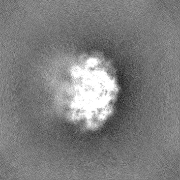
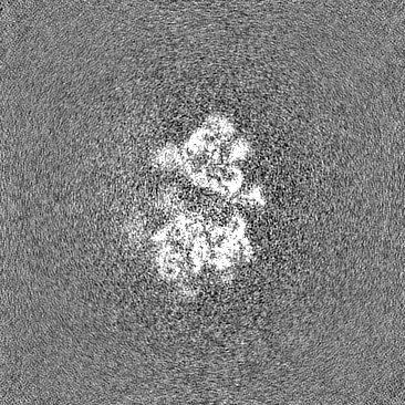
-Half map: Class C2 CP45 50S Ribosomal Intermediate - Half Map 2
| File | emd_43846_half_map_1.map | ||||||||||||
|---|---|---|---|---|---|---|---|---|---|---|---|---|---|
| Annotation | Class C2 CP45 50S Ribosomal Intermediate - Half Map 2 | ||||||||||||
| Projections & Slices |
| ||||||||||||
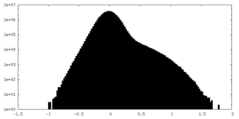
| Density Histograms |
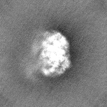


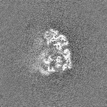
-Half map: Class C2 CP45 50S Ribosomal Intermediate - Half Map 1
| File | emd_43846_half_map_2.map | ||||||||||||
|---|---|---|---|---|---|---|---|---|---|---|---|---|---|
| Annotation | Class C2 CP45 50S Ribosomal Intermediate - Half Map 1 | ||||||||||||
| Projections & Slices |
| ||||||||||||
| Density Histograms |


- Sample components
-Entire : CP45 iSAT 50S ribosomal subunit assembly intermediate
| Entire | Name: CP45 iSAT 50S ribosomal subunit assembly intermediate |
|---|---|
| Components |
|
-Supramolecule #1: CP45 iSAT 50S ribosomal subunit assembly intermediate
| Supramolecule | Name: CP45 iSAT 50S ribosomal subunit assembly intermediate / type: complex / ID: 1 / Parent: 0 |
|---|---|
| Source (natural) | Organism: |
-Experimental details
-Structure determination
| Method | cryo EM |
|---|---|
 Processing Processing | single particle reconstruction |
| Aggregation state | particle |
-Sample preparation
| Buffer | pH: 7.5 Component:
Details: Most of the sucrose was removed by spin concentration. | ||||||||||||||||||
|---|---|---|---|---|---|---|---|---|---|---|---|---|---|---|---|---|---|---|---|
| Grid | Model: Quantifoil R1.2/1.3 / Material: COPPER / Support film - Material: CARBON / Support film - topology: CONTINUOUS / Support film - Film thickness: 2 | ||||||||||||||||||
| Vitrification | Cryogen name: ETHANE / Chamber humidity: 90 % / Chamber temperature: 298 K / Instrument: FEI VITROBOT MARK IV / Details: 3 microliter of the sample was added.. | ||||||||||||||||||
| Details | The in vitro assembled large ribosomal subunit was purified by sucrose gradient and was spin-concentrated in a 100 kDa MW-cutoff filter. |
- Electron microscopy
| Microscope | FEI TALOS ARCTICA |
|---|---|
| Details | In order to account for highly preferred orientation of the specimen, data were acquired using tilts ranging at -20 degrees. |
| Image recording | Film or detector model: GATAN K2 SUMMIT (4k x 4k) / Detector mode: COUNTING / Number grids imaged: 1 / Number real images: 1599 / Average exposure time: 5.0 sec. / Average electron dose: 50.0 e/Å2 |
| Electron beam | Acceleration voltage: 200 kV / Electron source:  FIELD EMISSION GUN FIELD EMISSION GUN |
| Electron optics | C2 aperture diameter: 50.0 µm / Illumination mode: FLOOD BEAM / Imaging mode: BRIGHT FIELD / Nominal defocus max: 2.0 µm / Nominal defocus min: 1.0 µm / Nominal magnification: 36000 |
| Sample stage | Cooling holder cryogen: NITROGEN |
| Experimental equipment |  Model: Talos Arctica / Image courtesy: FEI Company |

