Movie
Movie Controller

[English] 日本語
 Yorodumi
- EMDB-43740: Cryo-EM structure of human tankyrase 2 SAM-PARP filament bound to... -
+ Open data

- Basic information
| Entry |  | |||||||||
|---|---|---|---|---|---|---|---|---|---|---|
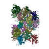
| Title | Cryo-EM structure of human tankyrase 2 SAM-PARP filament bound to compound, XAV (consensus map). | |||||||||
 Map data Map data | Local resolution filtered map | |||||||||
 Sample Sample |
| |||||||||
 Keywords Keywords | NAD+ ADP-ribosyltransferase / WNT signaling / inhibitor / SIGNALING PROTEIN / SIGNALING PROTEIN-INHIBITOR complex | |||||||||
| Function / homology |  Function and homology information Function and homology informationXAV939 stabilizes AXIN / positive regulation of telomere capping / NAD+ ADP-ribosyltransferase / protein auto-ADP-ribosylation / negative regulation of telomere maintenance via telomere lengthening / protein localization to chromosome, telomeric region / NAD+-protein-aspartate ADP-ribosyltransferase activity / protein poly-ADP-ribosylation / NAD+-protein-glutamate ADP-ribosyltransferase activity / NAD+-protein mono-ADP-ribosyltransferase activity ...XAV939 stabilizes AXIN / positive regulation of telomere capping / NAD+ ADP-ribosyltransferase / protein auto-ADP-ribosylation / negative regulation of telomere maintenance via telomere lengthening / protein localization to chromosome, telomeric region / NAD+-protein-aspartate ADP-ribosyltransferase activity / protein poly-ADP-ribosylation / NAD+-protein-glutamate ADP-ribosyltransferase activity / NAD+-protein mono-ADP-ribosyltransferase activity / pericentriolar material / Transferases; Glycosyltransferases; Pentosyltransferases / NAD+ poly-ADP-ribosyltransferase activity / carbohydrate transmembrane transporter activity / maltose binding / maltose transport / maltodextrin transmembrane transport / ATP-binding cassette (ABC) transporter complex, substrate-binding subunit-containing / positive regulation of telomere maintenance via telomerase / nucleotidyltransferase activity / TCF dependent signaling in response to WNT / Degradation of AXIN / Wnt signaling pathway / Regulation of PTEN stability and activity / protein polyubiquitination / nuclear envelope / positive regulation of canonical Wnt signaling pathway / outer membrane-bounded periplasmic space / chromosome, telomeric region / Ub-specific processing proteases / Golgi membrane / perinuclear region of cytoplasm / enzyme binding / metal ion binding / nucleus / cytoplasm / cytosol Similarity search - Function | |||||||||
| Biological species |  Homo sapiens (human) Homo sapiens (human) | |||||||||
| Method | helical reconstruction / cryo EM / Resolution: 2.21 Å | |||||||||
 Authors Authors | Malone BF / Zimmerman JL / Dow LE / Hite RK | |||||||||
| Funding support | 1 items
| |||||||||
 Citation Citation | Journal: bioRxiv / Year: 2025 Title: A potent and selective TNKS2 inhibitor for tumor-selective WNT suppression. Authors: Jill Zimmerman / Brandon F Malone / Efrat Finkin-Groner / Shan Sun / Rui Liang / Miguel Foronda / Emma M Schatoff / Elizabeth Granowsky / Sukanya Goswami / Alyna Katti / Benjamin Leach / ...Authors: Jill Zimmerman / Brandon F Malone / Efrat Finkin-Groner / Shan Sun / Rui Liang / Miguel Foronda / Emma M Schatoff / Elizabeth Granowsky / Sukanya Goswami / Alyna Katti / Benjamin Leach / Heather Alcorn / Tuomas Tammela / Yoshiyuki Fukase / Tanweer Khan / David J Huggins / John Ginn / Nigel Liverton / Richard K Hite / Lukas E Dow /  Abstract: Hyperactive WNT signaling is a potent cancer driver, but clinical translation of WNT inhibitors has been hampered by on-target toxicities. WNT signaling can be constrained through inhibition of the ...Hyperactive WNT signaling is a potent cancer driver, but clinical translation of WNT inhibitors has been hampered by on-target toxicities. WNT signaling can be constrained through inhibition of the PARP family enzymes Tankyrase 1 (TNKS1) and Tankyrase 2 (TNKS2), however, existing TNKS inhibitors suppress WNT signaling in both tumor and healthy tissues. In this study, we show that the loss of chromosome 8p that occurs in approximately half of advanced epithelial malignancies, creates a collateral vulnerability that enables tumor-selective inhibition of Tankyrase activity. 8p loss depletes expression of TNKS1 and creates a tumor-specific dependency on the functionally redundant TNKS2 protein. Through structure-guided drug design, we identify a first-in-class TNKS2-selective inhibitor that can drive selective WNT inhibition in TNKS1-deficient oncogenic cell and organoid models. This work demonstrates a targetable vulnerability in multiple cancer types, providing a new approach to potent and selective WNT-targeted therapies. | |||||||||
| History |
|
- Structure visualization
| Supplemental images |
|---|

- Downloads & links
-EMDB archive
| Map data | emd_43740.map.gz | 8.5 MB | EMDB map data format | |
|---|---|---|---|---|
| Header (meta data) | emd-43740-v30.xmlemd-43740.xml | 24.2 KB 24.2 KB | Display Display | EMDB header |
| FSC (resolution estimation) | emd_43740_fsc.xml | 12.6 KB | Display | FSC data file |
| Images |  emd_43740.png emd_43740.png | 83.9 KB | ||
| Filedesc metadata | emd-43740.cif.gz | 7.1 KB | ||
| Others | emd_43740_additional_1.map.gzemd_43740_additional_2.map.gzemd_43740_half_map_1.map.gzemd_43740_half_map_2.map.gz | 203.8 MB 108.2 MB 200.4 MB 200.4 MB | ||
| Archive directory |  http://ftp.pdbj.org/pub/emdb/structures/EMD-43740ftp://ftp.pdbj.org/pub/emdb/structures/EMD-43740 http://ftp.pdbj.org/pub/emdb/structures/EMD-43740ftp://ftp.pdbj.org/pub/emdb/structures/EMD-43740 | HTTPS FTP |
-Related structure data
| Related structure data |  8w27MC  8w23C  8w25C  8w28C  8w2tC  8w2uC C: citing same article ( M: atomic model generated by this map |
|---|---|
| Similar structure data |


-Links
| EMDB pages | EMDB (EBI/PDBe) / EMDataResource |
|---|---|
| Related items in Molecule of the Month |


-Map
| File | Download / File: emd_43740.map.gz / Format: CCP4 / Size: 216 MB / Type: IMAGE STORED AS FLOATING POINT NUMBER (4 BYTES) | ||||||||||||||||||||||||||||||||||||
|---|---|---|---|---|---|---|---|---|---|---|---|---|---|---|---|---|---|---|---|---|---|---|---|---|---|---|---|---|---|---|---|---|---|---|---|---|---|
| Annotation | Local resolution filtered map | ||||||||||||||||||||||||||||||||||||
| Projections & slices | Image control
Images are generated by Spider. | ||||||||||||||||||||||||||||||||||||
| Voxel size | X=Y=Z: 0.826 Å | ||||||||||||||||||||||||||||||||||||
| Density |
| ||||||||||||||||||||||||||||||||||||
| Symmetry | Space group: 1 | ||||||||||||||||||||||||||||||||||||
| Details | EMDB XML:
|
 Z (Sec.)
 Y (Row.)
 X (Col.)


-Supplemental data
-Additional map: B-factor sharpened map
| File | emd_43740_additional_1.map | ||||||||||||
|---|---|---|---|---|---|---|---|---|---|---|---|---|---|
| Annotation | B-factor sharpened map | ||||||||||||
| Projections & Slices |
| ||||||||||||
| Density Histograms |


-Additional map: Raw map
| File | emd_43740_additional_2.map | ||||||||||||
|---|---|---|---|---|---|---|---|---|---|---|---|---|---|
| Annotation | Raw map | ||||||||||||
| Projections & Slices |
| ||||||||||||
| Density Histograms |


-Half map: Half map A
| File | emd_43740_half_map_1.map | ||||||||||||
|---|---|---|---|---|---|---|---|---|---|---|---|---|---|
| Annotation | Half map A | ||||||||||||
| Projections & Slices |
| ||||||||||||
| Density Histograms |


-Half map: Half map B
| File | emd_43740_half_map_2.map | ||||||||||||
|---|---|---|---|---|---|---|---|---|---|---|---|---|---|
| Annotation | Half map B | ||||||||||||
| Projections & Slices |
| ||||||||||||
| Density Histograms |


- Sample components
-Entire : Tankyrase 2 SAM-PARP filament bound to compound XAV.
| Entire | Name: Tankyrase 2 SAM-PARP filament bound to compound XAV. |
|---|---|
| Components |
|
-Supramolecule #1: Tankyrase 2 SAM-PARP filament bound to compound XAV.
| Supramolecule | Name: Tankyrase 2 SAM-PARP filament bound to compound XAV. / type: complex / ID: 1 / Parent: 0 / Macromolecule list: #1 |
|---|---|
| Source (natural) | Organism: Homo sapiens (human) |
-Macromolecule #1: Maltose/maltodextrin-binding periplasmic protein,Poly [ADP-ribose...
| Macromolecule | Name: Maltose/maltodextrin-binding periplasmic protein,Poly [ADP-ribose] polymerase tankyrase-2 type: protein_or_peptide / ID: 1 Details: Tankyrase 2 fused to N-terminal twin-strep,Tankyrase 2 fused to N-terminal twin-strep Number of copies: 20 / Enantiomer: LEVO / EC number: NAD+ ADP-ribosyltransferase |
|---|---|
| Source (natural) | Organism: Homo sapiens (human) |
| Molecular weight | Theoretical: 80.614008 KDa |
| Recombinant expression | Organism:   Spodoptera frugiperda (fall armyworm) Spodoptera frugiperda (fall armyworm) |
| Sequence | String: MSAWSHPQFE KGGGSGGGSG GSAWSHPQFE KTGSMGIEEG KLVIWINGDK GYNGLAEVGK KFEKDTGIKV TVEHPDKLEE KFPQVAATG DGPDIIFWAH DRFGGYAQSG LLAEITPDKA FQDKLYPFTW DAVRYNGKLI AYPIAVEALS LIYNKDLLPN P PKTWEEIP ...String: MSAWSHPQFE KGGGSGGGSG GSAWSHPQFE KTGSMGIEEG KLVIWINGDK GYNGLAEVGK KFEKDTGIKV TVEHPDKLEE KFPQVAATG DGPDIIFWAH DRFGGYAQSG LLAEITPDKA FQDKLYPFTW DAVRYNGKLI AYPIAVEALS LIYNKDLLPN P PKTWEEIP ALDKELKAKG KSALMFNLQE PYFTWPLIAA DGGYAFKYEN GKYDIKDVGV DNAGAKAGLT FLVDLIKNKH MN ADTDYSI AEAAFNKGET AMTINGPWAW SNIDTSKVNY GVTVLPTFKG QPSKPFVGVL SAGINAASPN KELAKEFLEN YLL TDEGLE AVNKDKPLGA VALKSYEEEL AKDPRIAATM ENAQKGEIMP NIPQMSAFWY AVRTAVINAA SGRQTVDEAL KDAQ TELEL EVLFQGPELS SVVSSSGTEG ASSLEKKEVP GVDFSITQFV RNLGLEHLMD IFEREQITLD VLVEMGHKEL KEIGI NAYG HRHKLIKGVE RLISGQQGLN PYLTLNTSGS GTILIDLSPD DKEFQSVEEE MQSTVREHRD GGHAGGIFNR YNILKI QKV CNKKLWERYT HRRKEVSEEN HNHANERMLF HGSPFVNAII HKGFDERHAY IGGMFGAGIY FAENSSKSNQ YVYGIGG GT GCPVHKDRSC YICHRQLLFC RVTLGKSFLQ FSAMKMAHSP PGHHSVTGRP SVNGLALAEY VIYRGEQAYP EYLITYQI M RPEGMVDG UniProtKB: Maltose/maltodextrin-binding periplasmic protein, Poly [ADP-ribose] polymerase tankyrase-2 |
-Macromolecule #2: 2-[4-(trifluoromethyl)phenyl]-7,8-dihydro-5H-thiopyrano[4,3-d]pyr...
| Macromolecule | Name: 2-[4-(trifluoromethyl)phenyl]-7,8-dihydro-5H-thiopyrano[4,3-d]pyrimidin-4-ol type: ligand / ID: 2 / Number of copies: 20 / Formula: XAV |
|---|---|
| Molecular weight | Theoretical: 312.31 Da |
| Chemical component information | 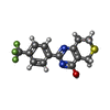 ChemComp-XAV: |
-Macromolecule #3: ZINC ION
| Macromolecule | Name: ZINC ION / type: ligand / ID: 3 / Number of copies: 20 / Formula: ZN |
|---|---|
| Molecular weight | Theoretical: 65.409 Da |
-Experimental details
-Structure determination
| Method | cryo EM |
|---|---|
 Processing Processing | helical reconstruction |
| Aggregation state | filament |
-Sample preparation
| Concentration | 1.3 mg/mL | ||||||||||||||||||
|---|---|---|---|---|---|---|---|---|---|---|---|---|---|---|---|---|---|---|---|
| Buffer | pH: 7.5 Component:
Details: Sample was purified and concentrated in the above buffer but exchanged prior to vitrification to a low-salt (minimal) buffer composed of 20 millimolar HEPES pH 7.5. Sample buffer was ...Details: Sample was purified and concentrated in the above buffer but exchanged prior to vitrification to a low-salt (minimal) buffer composed of 20 millimolar HEPES pH 7.5. Sample buffer was exchanged on grid via manual side-blotting. | ||||||||||||||||||
| Vitrification | Cryogen name: ETHANE / Chamber humidity: 95 % / Chamber temperature: 283 K / Instrument: FEI VITROBOT MARK IV |
- Electron microscopy
| Microscope | TFS KRIOS |
|---|---|
| Image recording | Film or detector model: GATAN K3 (6k x 4k) / Average electron dose: 55.7 e/Å2 |
| Electron beam | Acceleration voltage: 300 kV / Electron source:  FIELD EMISSION GUN FIELD EMISSION GUN |
| Electron optics | Illumination mode: FLOOD BEAM / Imaging mode: BRIGHT FIELD / Cs: 2.7 mm / Nominal defocus max: 3.0 µm / Nominal defocus min: 0.8 µm / Nominal magnification: 29000 |
| Sample stage | Specimen holder model: FEI TITAN KRIOS AUTOGRID HOLDER / Cooling holder cryogen: NITROGEN |
| Experimental equipment |  Model: Titan Krios / Image courtesy: FEI Company |