 Movie
Movie Controller


+ Open data

- Basic information
| Entry |  | |||||||||
|---|---|---|---|---|---|---|---|---|---|---|
| Title | BsaXI -- Type IIB R-M system | |||||||||
 Map data Map data | cryosparc2_P35_J607, app BsaXI | |||||||||
 Sample Sample |
| |||||||||
 Keywords Keywords | R-M system / restriction nucleases / Type IIB / protein-DNA complex / BsaXI. / ANTIMICROBIAL PROTEIN | |||||||||
| Function / homology | : / Type I restriction modification DNA specificity domain / Type I restriction modification DNA specificity domain superfamily / Type I restriction modification DNA specificity domain / DNA restriction-modification system / DNA binding / Uncharacterized protein / :  Function and homology information Function and homology information | |||||||||
| Biological species |   Geobacillus stearothermophilus (bacteria) Geobacillus stearothermophilus (bacteria) | |||||||||
| Method | single particle reconstruction / cryo EM / Resolution: 3.26 Å | |||||||||
 Authors Authors | Shen BW / Stoddard BL | |||||||||
| Funding support |  United States, 1 items United States, 1 items
| |||||||||
 Citation Citation | Journal: To Be Published Title: Atomic insights into the mechanism of DNA recognition and methylation by type IIB restriction-modification system: CryoEM structures of DNA free and cognate DNA bound BsaXI. Authors: Shen BW / Stoddard BL | |||||||||
| History |
|
- Structure visualization
| Supplemental images |
|---|

- Downloads & links
-EMDB archive
| Map data | emd_43711.map.gz | 108.7 MB | EMDB map data format | |
|---|---|---|---|---|
| Header (meta data) | emd-43711-v30.xmlemd-43711.xml | 22.2 KB 22.2 KB | Display Display | EMDB header |
| FSC (resolution estimation) | emd_43711_fsc.xml | 12.7 KB | Display | FSC data file |
| Images |  emd_43711.png emd_43711.png | 111.3 KB | ||
| Filedesc metadata | emd-43711.cif.gz | 7.3 KB | ||
| Others | emd_43711_half_map_1.map.gzemd_43711_half_map_2.map.gz | 200.2 MB 200.2 MB | ||
| Archive directory |  http://ftp.pdbj.org/pub/emdb/structures/EMD-43711ftp://ftp.pdbj.org/pub/emdb/structures/EMD-43711 http://ftp.pdbj.org/pub/emdb/structures/EMD-43711ftp://ftp.pdbj.org/pub/emdb/structures/EMD-43711 | HTTPS FTP |
-Related structure data
| Related structure data | 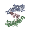 8w0pMC M: atomic model generated by this map C: citing same article ( |
|---|---|
| Similar structure data |
-Links
| EMDB pages | EMDB (EBI/PDBe) / EMDataResource |
|---|
-Map
| File | Download / File: emd_43711.map.gz / Format: CCP4 / Size: 216 MB / Type: IMAGE STORED AS FLOATING POINT NUMBER (4 BYTES) | ||||||||||||||||||||||||||||||||||||
|---|---|---|---|---|---|---|---|---|---|---|---|---|---|---|---|---|---|---|---|---|---|---|---|---|---|---|---|---|---|---|---|---|---|---|---|---|---|
| Annotation | cryosparc2_P35_J607, app BsaXI | ||||||||||||||||||||||||||||||||||||
| Projections & slices | Image control
Images are generated by Spider. | ||||||||||||||||||||||||||||||||||||
| Voxel size | X=Y=Z: 1.079 Å | ||||||||||||||||||||||||||||||||||||
| Density |
| ||||||||||||||||||||||||||||||||||||
| Symmetry | Space group: 1 | ||||||||||||||||||||||||||||||||||||
| Details | EMDB XML:
|
 Z (Sec.)
 Y (Row.)
 X (Col.)


-Supplemental data
-Half map: apoBsaXI; half map B CS P35 J607 B
| File | emd_43711_half_map_1.map | ||||||||||||
|---|---|---|---|---|---|---|---|---|---|---|---|---|---|
| Annotation | apoBsaXI; half_map_B CS_P35_J607_B | ||||||||||||
| Projections & Slices |
| ||||||||||||
| Density Histograms |


-Half map: apoBsaXI, half map A CS P35 607 A
| File | emd_43711_half_map_2.map | ||||||||||||
|---|---|---|---|---|---|---|---|---|---|---|---|---|---|
| Annotation | apoBsaXI, half_map_A CS_P35_607_A | ||||||||||||
| Projections & Slices |
| ||||||||||||
| Density Histograms |


- Sample components
-Entire : heterotrimers of RM fusion and S subunit
| Entire | Name: heterotrimers of RM fusion and S subunit |
|---|---|
| Components |
|
-Supramolecule #1: heterotrimers of RM fusion and S subunit
| Supramolecule | Name: heterotrimers of RM fusion and S subunit / type: complex / ID: 1 / Parent: 0 / Macromolecule list: #1-#2 Details: reconstituted RM and S subunits of type IIB R-M systems. |
|---|---|
| Source (natural) | Organism: Geobacillus stearothermophilus (bacteria) / Strain: Cpw230 |
| Molecular weight | Theoretical: 590 KDa |
-Macromolecule #1: RM.BsaXI
| Macromolecule | Name: RM.BsaXI / type: protein_or_peptide / ID: 1 / Details: RM fusion protein / Number of copies: 2 / Enantiomer: LEVO EC number: site-specific DNA-methyltransferase (adenine-specific) |
|---|---|
| Source (natural) | Organism: Geobacillus stearothermophilus (bacteria) / Strain: Cpw230 |
| Molecular weight | Theoretical: 107.195234 KDa |
| Recombinant expression | Organism: |
| Sequence | String: MKNWQRIVEA KLEQQKHKVA EISLENGTVN YSKKIKHNRN LKALTGDEEI VRAFLIDRLV NELDYKPEYL ETEKEYTIKG GHSKINPRV DVLVKDDKGN PFFFIEVKAP NKFEEDKDEI EGQLFALAQA EERDFKTKVK YLVYYTVELI DDEIVDRAII I DFEKYPTY ...String: MKNWQRIVEA KLEQQKHKVA EISLENGTVN YSKKIKHNRN LKALTGDEEI VRAFLIDRLV NELDYKPEYL ETEKEYTIKG GHSKINPRV DVLVKDDKGN PFFFIEVKAP NKFEEDKDEI EGQLFALAQA EERDFKTKVK YLVYYTVELI DDEIVDRAII I DFEKYPTY TDWSNGGFIS TGTELTAGYG EPKKQPLIKG HEKYDLRVRI DREEIEGLGR NLHNVLWGGG GTNDSEIFYS LV NIILAKI QDEYEKEDGQ EYDFQVYQYG DNVESPQKLF DRINALYKRA LREQLNVTDE QKIAEDNVIN RNKFPLNKLV YTV QALESL SFLEGRNSLD GKDILGDFFE SIIRDGFKQT KGQFFTPTPI VKFILYALQL DKLAIDRLNN DRELPLIIDP SAGS GTFLI EAMKLITKEV KYKQNHKVKS SRQITKRFEE LFMPDHNENK WAREYLYGCE INFDLGTASK VNMILHGDGS ANIFV QDGL LPFRFYVKET SPNYLETASP DALYGDKEVN GKFDVVVSNP PFSVDLDTQT QREVRNAFLF GDKKNSENLF IERYYQ LLK EGGRLGVVLP ESVFDTTENK YIRLFIFKYF KVKAVVSLPQ VTFEPFTSTK TSLLFAQKKT KEEVEQWNEL WDKYGKE WS LLKTRINDYF SYFVKGRPLN KKWAPDVVKD IQEGNEDNIR KNIFRFLKDH IKEEDKNLEI KDLLIKYAEE ISSISKHE K ETDVFGFYNA WWVFGEVAKE LDYPIFMAEA ENVGYKRTKK GEKPMPNDLY DLEYAPSTLD CEKVLSSFDI EINALEASK TKLSVEKGLL EEKLKDKEDK ENEKIQKRLN KISELLETIE NQLDSIRSKK LEVEGILEKY YENNKLKEEY SERDDEELIN HFKHGVLYQ YRSEDILLRN KTVHKILDEI RQGVIWD UniProtKB: UNIPROTKB: A0A4D7QEP1 |
-Macromolecule #2: S.BsaXI
| Macromolecule | Name: S.BsaXI / type: protein_or_peptide / ID: 2 / Number of copies: 1 / Enantiomer: LEVO |
|---|---|
| Source (natural) | Organism: Geobacillus stearothermophilus (bacteria) / Strain: Cpw230 |
| Molecular weight | Theoretical: 55.038824 KDa |
| Recombinant expression | Organism: |
| Sequence | String: MGLIQRRNFS TFASEPSVRF DFNYMKSVTP TTEEYYTYKS LFEVVPSTVP TLDESEPFKY AEIGHVSKNG EVFPVTLSFE DRDELNEDL FKKIEKGDIF LPERGNILIS AIRPYLNKIV LIKEDDKTDI YFTKAFIQIK PLINSRILYY ALRTIFSEKI N AVSRQGKG ...String: MGLIQRRNFS TFASEPSVRF DFNYMKSVTP TTEEYYTYKS LFEVVPSTVP TLDESEPFKY AEIGHVSKNG EVFPVTLSFE DRDELNEDL FKKIEKGDIF LPERGNILIS AIRPYLNKIV LIKEDDKTDI YFTKAFIQIK PLINSRILYY ALRTIFSEKI N AVSRQGKG YPTLKEDDLK TIQFSKKVID NLLAKEEELI SNIDALEKDI KELKSIQRSK KEIVDEVFSS HFNINMVELM AL DSQRRVD VGLSSISSLN STIRYSYRWN KMKLIQKYLY RDIDCIEPLG KYILSSNNGW SPESVVGGEG IPILGQEHLE FDG VLNVSP TKATTKTKNN MENFFIQEGD LFISRGNTVD LVGLACVVET EVTEDIIYPD LYIRLKIDEK VIHKKYLALL FNSF FGRLY FKYVSKGKNQ TMVKISSNEL LNYYLPIPPM EEQLEIVGKI EEQIGAQNEI EKQIEEKRNQ IRVIIEETAR S UniProtKB: Uncharacterized protein |
-Macromolecule #3: S-ADENOSYLMETHIONINE
| Macromolecule | Name: S-ADENOSYLMETHIONINE / type: ligand / ID: 3 / Number of copies: 2 / Formula: SAM |
|---|---|
| Molecular weight | Theoretical: 398.437 Da |
| Chemical component information |  ChemComp-SAM: |
-Experimental details
-Structure determination
| Method | cryo EM |
|---|---|
 Processing Processing | single particle reconstruction |
| Aggregation state | particle |
-Sample preparation
| Concentration | 0.35 mg/mL |
|---|---|
| Buffer | pH: 8 / Details: 20 mM TrisHCl, ph 8.0, 2 mM CaCl2, 150 mM NaCl |
| Grid | Model: Quantifoil R1.2/1.3 / Material: COPPER / Mesh: 300 / Support film - Material: CARBON / Support film - topology: HOLEY / Pretreatment - Type: GLOW DISCHARGE / Pretreatment - Time: 30 sec. / Pretreatment - Atmosphere: AIR |
| Vitrification | Cryogen name: ETHANE / Chamber humidity: 95 % / Chamber temperature: 298 K / Instrument: FEI VITROBOT MARK IV |
| Details | Monodisperse |
- Electron microscopy
| Microscope | FEI TITAN KRIOS |
|---|---|
| Image recording | Film or detector model: GATAN K2 SUMMIT (4k x 4k) / Detector mode: COUNTING / Digitization - Frames/image: 2-50 / Number grids imaged: 1 / Number real images: 6227 / Average exposure time: 2.0 sec. / Average electron dose: 40.0 e/Å2 |
| Electron beam | Acceleration voltage: 300 kV / Electron source:  FIELD EMISSION GUN FIELD EMISSION GUN |
| Electron optics | C2 aperture diameter: 70.0 µm / Illumination mode: FLOOD BEAM / Imaging mode: BRIGHT FIELD / Cs: 2.7 mm / Nominal defocus max: 5.0 µm / Nominal defocus min: 1.2 µm |
| Experimental equipment |  Model: Titan Krios / Image courtesy: FEI Company |

