 ムービー
ムービー コントローラー


+ データを開く

- 基本情報
基本情報
| 登録情報 |  | |||||||||
|---|---|---|---|---|---|---|---|---|---|---|
| タイトル | Tomogram 7 - Thickness measurement | |||||||||
 マップデータ マップデータ | ||||||||||
 試料 試料 |
| |||||||||
 キーワード キーワード | ER-HOXB8 cells / neutrophils / cytoplasm / RIBOSOME | |||||||||
| 生物種 |  | |||||||||
| 手法 | 電子線トモグラフィー法 / クライオ電子顕微鏡法 | |||||||||
 データ登録者 データ登録者 | Elferich J / Kong L / Zottig X / Grigorieff N | |||||||||
| 資金援助 |  米国, 1件 米国, 1件
| |||||||||
 引用 引用 | ジャーナル: Elife / 年: 2024 タイトル: CTFFIND5 provides improved insight into quality, tilt, and thickness of TEM samples. 著者: Johannes Elferich / Lingli Kong / Ximena Zottig / Nikolaus Grigorieff / 要旨: Images taken by transmission electron microscopes are usually affected by lens aberrations and image defocus, among other factors. These distortions can be modeled in reciprocal space using the ...Images taken by transmission electron microscopes are usually affected by lens aberrations and image defocus, among other factors. These distortions can be modeled in reciprocal space using the contrast transfer function (CTF). Accurate estimation and correction of the CTF is essential for restoring the high-resolution signal in cryogenic electron microscopy (cryoEM). Previously, we described the implementation of algorithms for this task in the TEM software package (Grant et al., 2018). Here we show that taking sample characteristics, such as thickness and tilt, into account can improve CTF estimation. This is particularly important when imaging cellular samples, where measurement of sample thickness and geometry derived from accurate modeling of the Thon ring pattern helps judging the quality of the sample. This improved CTF estimation has been implemented in CTFFIND5, a new version of the TEM program CTFFIND. We evaluated the accuracy of these estimates using images of tilted aquaporin crystals and eukaryotic cells thinned by focused ion beam milling. We estimate that with micrographs of sufficient quality CTFFIND5 can measure sample tilt with an accuracy of 3° and sample thickness with an accuracy of 5 nm. #1: ジャーナル: Elife / 年: 2024タイトル: CTFFIND5 provides improved insight into quality, tilt and thickness of TEM sample 著者: Elferich J / Kong L / Zottig X / Grigorieff N | |||||||||
| 履歴 |
|
- 構造の表示
構造の表示
| 添付画像 |
|---|
- ダウンロードとリンク
-EMDBアーカイブ
| マップデータ | emd_43429.map.gz | 423.1 MB |  EMDBマップデータ形式 EMDBマップデータ形式 | |
|---|---|---|---|---|
| ヘッダ (付随情報) | emd-43429-v30.xmlemd-43429.xml | 8.5 KB 8.5 KB | 表示 表示 | EMDBヘッダ |
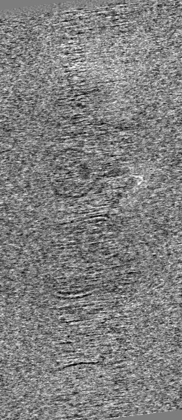
| 画像 |  emd_43429.png emd_43429.png | 126.1 KB | ||
| Filedesc metadata | emd-43429.cif.gz | 3.5 KB | ||
| アーカイブディレクトリ |  http://ftp.pdbj.org/pub/emdb/structures/EMD-43429ftp://ftp.pdbj.org/pub/emdb/structures/EMD-43429 http://ftp.pdbj.org/pub/emdb/structures/EMD-43429ftp://ftp.pdbj.org/pub/emdb/structures/EMD-43429 | HTTPS FTP |
-検証レポート
| 文書・要旨 | emd_43429_validation.pdf.gz | 607.1 KB | 表示 | EMDB検証レポート |
|---|---|---|---|---|
| 文書・詳細版 | emd_43429_full_validation.pdf.gz | 606.7 KB | 表示 | |
| XML形式データ | emd_43429_validation.xml.gz | 2.5 KB | 表示 | |
| CIF形式データ | emd_43429_validation.cif.gz | 3.1 KB | 表示 | |
| アーカイブディレクトリ | https://ftp.pdbj.org/pub/emdb/validation_reports/EMD-43429ftp://ftp.pdbj.org/pub/emdb/validation_reports/EMD-43429 | HTTPS FTP |
-関連構造データ


-リンク
| EMDBのページ | EMDB (EBI/PDBe) / EMDataResource |
|---|
-マップ
| ファイル | ダウンロード / ファイル: emd_43429.map.gz / 形式: CCP4 / 大きさ: 1.7 GB / タイプ: IMAGE STORED AS SIGNED INTEGER (2 BYTES) | ||||||||||||||||||||||||||||||||
|---|---|---|---|---|---|---|---|---|---|---|---|---|---|---|---|---|---|---|---|---|---|---|---|---|---|---|---|---|---|---|---|---|---|
| 投影像・断面図 | 画像のコントロール
画像は Spider により作成 これらの図は立方格子座標系で作成されたものです | ||||||||||||||||||||||||||||||||
| ボクセルのサイズ | X=Y=Z: 8.349 Å | ||||||||||||||||||||||||||||||||
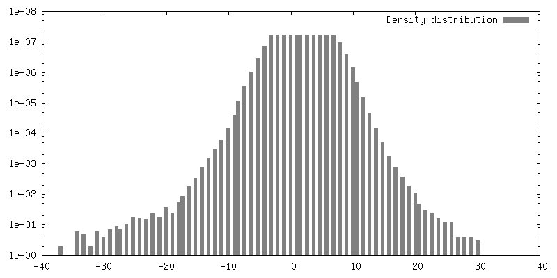
| 密度 |
| ||||||||||||||||||||||||||||||||
| 対称性 | 空間群: 1 | ||||||||||||||||||||||||||||||||
| 詳細 | EMDB XML:
|
 Z (Sec.)
 Y (Row.)
 X (Col.)


-添付データ
- 試料の構成要素
-全体 : ER-HOXB8 sample prepared by FIB_SEM milling
| 全体 | 名称: ER-HOXB8 sample prepared by FIB_SEM milling |
|---|---|
| 要素 |
|
-超分子 #1: ER-HOXB8 sample prepared by FIB_SEM milling
| 超分子 | 名称: ER-HOXB8 sample prepared by FIB_SEM milling / タイプ: cell / ID: 1 / 親要素: 0 |
|---|---|
| 由来(天然) | 生物種: |
-実験情報
-構造解析
| 手法 | クライオ電子顕微鏡法 |
|---|---|
 解析 解析 | 電子線トモグラフィー法 |
| 試料の集合状態 | cell |
-試料調製
| 緩衝液 | pH: 7.5 |
|---|---|
| 凍結 | 凍結剤: ETHANE / 装置: LEICA PLUNGER |
| 切片作成 | 集束イオンビーム - 装置: OTHER / 集束イオンビーム - イオン: OTHER / 集束イオンビーム - 電圧: 30 / 集束イオンビーム - 電流: 0.03 / 集束イオンビーム - 時間: 60 / 集束イオンビーム - 温度: 273 K / 集束イオンビーム - Initial thickness: 1000 / 集束イオンビーム - 最終 厚さ: 200 集束イオンビーム - 詳細: The value given for _em_focused_ion_beam.instrument is Aquilos 2. This is not in a list of allowed values {'OTHER', 'DB235'} so OTHER is written into the XML file. |
- 電子顕微鏡法
| 顕微鏡 | FEI TITAN KRIOS |
|---|---|
| 撮影 | フィルム・検出器のモデル: GATAN K3 (6k x 4k) / 平均電子線量: 3.0 e/Å2 |
| 電子線 | 加速電圧: 300 kV / 電子線源:  FIELD EMISSION GUN FIELD EMISSION GUN |
| 電子光学系 | 照射モード: FLOOD BEAM / 撮影モード: BRIGHT FIELD / Cs: 2.7 mm / 最大 デフォーカス(公称値): 8.0 µm / 最小 デフォーカス(公称値): 6.0 µm |
| 実験機器 |  モデル: Titan Krios / 画像提供: FEI Company |
-画像解析
| 最終 再構成 | 使用した粒子像数: 34 |
|---|
