 Movie
Movie Controller


+ Open data

- Basic information
| Entry |  | |||||||||
|---|---|---|---|---|---|---|---|---|---|---|
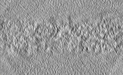
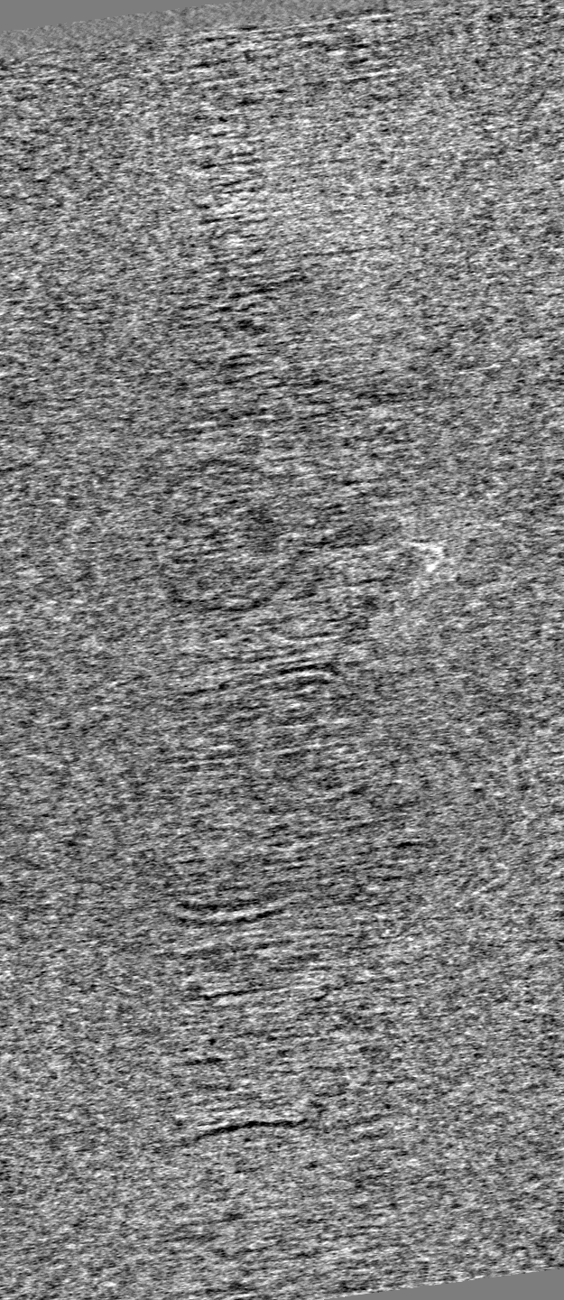
| Title | Tomogram 7 - Thickness measurement | |||||||||
 Map data Map data | ||||||||||
 Sample Sample |
| |||||||||
 Keywords Keywords | ER-HOXB8 cells / neutrophils / cytoplasm / RIBOSOME | |||||||||
| Biological species |  | |||||||||
| Method | electron tomography / cryo EM | |||||||||
 Authors Authors | Elferich J / Kong L / Zottig X / Grigorieff N | |||||||||
| Funding support |  United States, 1 items United States, 1 items
| |||||||||
 Citation Citation | Journal: Elife / Year: 2024 Title: CTFFIND5 provides improved insight into quality, tilt, and thickness of TEM samples. Authors: Johannes Elferich / Lingli Kong / Ximena Zottig / Nikolaus Grigorieff / Abstract: Images taken by transmission electron microscopes are usually affected by lens aberrations and image defocus, among other factors. These distortions can be modeled in reciprocal space using the ...Images taken by transmission electron microscopes are usually affected by lens aberrations and image defocus, among other factors. These distortions can be modeled in reciprocal space using the contrast transfer function (CTF). Accurate estimation and correction of the CTF is essential for restoring the high-resolution signal in cryogenic electron microscopy (cryoEM). Previously, we described the implementation of algorithms for this task in the TEM software package (Grant et al., 2018). Here we show that taking sample characteristics, such as thickness and tilt, into account can improve CTF estimation. This is particularly important when imaging cellular samples, where measurement of sample thickness and geometry derived from accurate modeling of the Thon ring pattern helps judging the quality of the sample. This improved CTF estimation has been implemented in CTFFIND5, a new version of the TEM program CTFFIND. We evaluated the accuracy of these estimates using images of tilted aquaporin crystals and eukaryotic cells thinned by focused ion beam milling. We estimate that with micrographs of sufficient quality CTFFIND5 can measure sample tilt with an accuracy of 3° and sample thickness with an accuracy of 5 nm. #1: Journal: Elife / Year: 2024Title: CTFFIND5 provides improved insight into quality, tilt and thickness of TEM sample Authors: Elferich J / Kong L / Zottig X / Grigorieff N | |||||||||
| History |
|
- Structure visualization
| Supplemental images |
|---|
- Downloads & links
-EMDB archive
| Map data | emd_43429.map.gz | 423.1 MB |  EMDB map data format EMDB map data format | |
|---|---|---|---|---|
| Header (meta data) | emd-43429-v30.xmlemd-43429.xml | 8.5 KB 8.5 KB | Display Display | EMDB header |
| Images |  emd_43429.png emd_43429.png | 126.1 KB | ||
| Filedesc metadata | emd-43429.cif.gz | 3.5 KB | ||
| Archive directory |  http://ftp.pdbj.org/pub/emdb/structures/EMD-43429ftp://ftp.pdbj.org/pub/emdb/structures/EMD-43429 http://ftp.pdbj.org/pub/emdb/structures/EMD-43429ftp://ftp.pdbj.org/pub/emdb/structures/EMD-43429 | HTTPS FTP |
-Validation report
| Summary document | emd_43429_validation.pdf.gz | 607.1 KB | Display | EMDB validaton report |
|---|---|---|---|---|
| Full document | emd_43429_full_validation.pdf.gz | 606.7 KB | Display | |
| Data in XML | emd_43429_validation.xml.gz | 2.5 KB | Display | |
| Data in CIF | emd_43429_validation.cif.gz | 3.1 KB | Display | |
| Arichive directory | https://ftp.pdbj.org/pub/emdb/validation_reports/EMD-43429ftp://ftp.pdbj.org/pub/emdb/validation_reports/EMD-43429 | HTTPS FTP |
-Related structure data


-Links
| EMDB pages | EMDB (EBI/PDBe) / EMDataResource |
|---|
-Map
| File | Download / File: emd_43429.map.gz / Format: CCP4 / Size: 1.7 GB / Type: IMAGE STORED AS SIGNED INTEGER (2 BYTES) | ||||||||||||||||||||||||||||||||
|---|---|---|---|---|---|---|---|---|---|---|---|---|---|---|---|---|---|---|---|---|---|---|---|---|---|---|---|---|---|---|---|---|---|
| Projections & slices | Image control
Images are generated by Spider. generated in cubic-lattice coordinate | ||||||||||||||||||||||||||||||||
| Voxel size | X=Y=Z: 8.349 Å | ||||||||||||||||||||||||||||||||
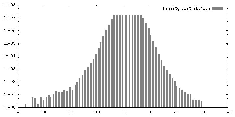
| Density |
| ||||||||||||||||||||||||||||||||
| Symmetry | Space group: 1 | ||||||||||||||||||||||||||||||||
| Details | EMDB XML:
|
 Z (Sec.)
 Y (Row.)
 X (Col.)


-Supplemental data
- Sample components
-Entire : ER-HOXB8 sample prepared by FIB_SEM milling
| Entire | Name: ER-HOXB8 sample prepared by FIB_SEM milling |
|---|---|
| Components |
|
-Supramolecule #1: ER-HOXB8 sample prepared by FIB_SEM milling
| Supramolecule | Name: ER-HOXB8 sample prepared by FIB_SEM milling / type: cell / ID: 1 / Parent: 0 |
|---|---|
| Source (natural) | Organism: |
-Experimental details
-Structure determination
| Method | cryo EM |
|---|---|
 Processing Processing | electron tomography |
| Aggregation state | cell |
-Sample preparation
| Buffer | pH: 7.5 |
|---|---|
| Vitrification | Cryogen name: ETHANE / Instrument: LEICA PLUNGER |
| Sectioning | Focused ion beam - Instrument: OTHER / Focused ion beam - Ion: OTHER / Focused ion beam - Voltage: 30 / Focused ion beam - Current: 0.03 / Focused ion beam - Duration: 60 / Focused ion beam - Temperature: 273 K / Focused ion beam - Initial thickness: 1000 / Focused ion beam - Final thickness: 200 Focused ion beam - Details: The value given for _em_focused_ion_beam.instrument is Aquilos 2. This is not in a list of allowed values {'OTHER', 'DB235'} so OTHER is written into the XML file. |
- Electron microscopy
| Microscope | FEI TITAN KRIOS |
|---|---|
| Image recording | Film or detector model: GATAN K3 (6k x 4k) / Average electron dose: 3.0 e/Å2 |
| Electron beam | Acceleration voltage: 300 kV / Electron source:  FIELD EMISSION GUN FIELD EMISSION GUN |
| Electron optics | Illumination mode: FLOOD BEAM / Imaging mode: BRIGHT FIELD / Cs: 2.7 mm / Nominal defocus max: 8.0 µm / Nominal defocus min: 6.0 µm |
| Experimental equipment |  Model: Titan Krios / Image courtesy: FEI Company |
-Image processing
| Final reconstruction | Number images used: 34 |
|---|
