 Movie
Movie Controller


+ Open data

- Basic information
| Entry |  | |||||||||
|---|---|---|---|---|---|---|---|---|---|---|
| Title | Cryo EM structure of a soybean CesA6 homotrimer | |||||||||
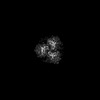
 Map data Map data | Single particle cryo EM map after non-uniform (C3) and local refinement in cryoSPARC. | |||||||||
 Sample Sample |
| |||||||||
 Keywords Keywords | Cellulose / glycosyltransferase / microfibril / TRANSFERASE | |||||||||
| Function / homology |  Function and homology information Function and homology informationplant-type primary cell wall biogenesis / cellulose synthase activity / cellulose synthase (UDP-forming) / cellulose synthase (UDP-forming) activity / cellulose biosynthetic process / cell wall organization / zinc ion binding / plasma membrane Similarity search - Function | |||||||||
| Biological species |  | |||||||||
| Method | single particle reconstruction / cryo EM / Resolution: 3.0 Å | |||||||||
 Authors Authors | Ho R / Palliniti P / Zimmer J | |||||||||
| Funding support |  United States, 1 items United States, 1 items
| |||||||||
 Citation Citation | Journal: Elife / Year: 2025 Title: Structure, function and assembly of soybean primary cell wall cellulose synthases. Authors: Ruoya Ho / Pallinti Purushotham / Louis F L Wilson / Yueping Wan / Jochen Zimmer / Abstract: Plant cell walls contain a meshwork of cellulose fibers embedded into a matrix of other carbohydrate and non-carbohydrate-based biopolymers. This composite material exhibits extraordinary properties, ...Plant cell walls contain a meshwork of cellulose fibers embedded into a matrix of other carbohydrate and non-carbohydrate-based biopolymers. This composite material exhibits extraordinary properties, from stretchable and pliable cell boundaries to solid protective shells. Cellulose, a linear glucose polymer, is synthesized and secreted across the plasma membrane by cellulose synthase (CesA), of which plants express multiple isoforms. Different subsets of CesA isoforms are necessary for primary and secondary cell wall biogenesis. Here, we structurally and functionally characterize the (soybean) primary cell wall CesAs CesA1, CesA3, and CesA6. The CesA isoforms exhibit robust in vitro catalytic activity. Cryo-electron microscopy analyses reveal their assembly into homotrimeric complexes in vitro in which each CesA protomer forms a cellulose-conducting transmembrane channel with a large lateral opening. Biochemical and co-purification analyses demonstrate that different CesA isoforms interact in vitro, leading to synergistic cellulose biosynthesis. Interactions between CesA trimers are only observed between different CesA isoforms and require the class-specific region (CSR). The CSR forms a hook-shaped extension of CesA's catalytic domain at the cytosolic water-lipid interface. Negative stain and cryo-electron microscopy analyses of mixtures of different CesA isoform trimers reveal their side-by-side arrangement into loose clusters. Our data suggest a model by which CesA homotrimers of different isoforms assemble into cellulose synthase complexes to synthesize and secrete multiple cellulose chains for microfibril formation. Inter-trimer interactions are mediated by fuzzy interactions between their CSR extensions. | |||||||||
| History |
|
- Structure visualization
| Supplemental images |
|---|

- Downloads & links
-EMDB archive
| Map data | emd_43245.map.gz | 230.3 MB | EMDB map data format | |
|---|---|---|---|---|
| Header (meta data) | emd-43245-v30.xmlemd-43245.xml | 17.8 KB 17.8 KB | Display Display | EMDB header |
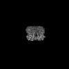
| Images |  emd_43245.png emd_43245.png | 179.8 KB | ||
| Filedesc metadata | emd-43245.cif.gz | 6.5 KB | ||
| Others | emd_43245_half_map_1.map.gzemd_43245_half_map_2.map.gz | 226.3 MB 226.3 MB | ||
| Archive directory |  http://ftp.pdbj.org/pub/emdb/structures/EMD-43245ftp://ftp.pdbj.org/pub/emdb/structures/EMD-43245 http://ftp.pdbj.org/pub/emdb/structures/EMD-43245ftp://ftp.pdbj.org/pub/emdb/structures/EMD-43245 | HTTPS FTP |
-Related structure data
| Related structure data |  8vi0MC  8vhtC  8vhzC M: atomic model generated by this map C: citing same article ( |
|---|---|
| Similar structure data |
-Links
| EMDB pages | EMDB (EBI/PDBe) / EMDataResource |
|---|---|
| Related items in Molecule of the Month |


-Map
| File | Download / File: emd_43245.map.gz / Format: CCP4 / Size: 244.1 MB / Type: IMAGE STORED AS FLOATING POINT NUMBER (4 BYTES) | ||||||||||||||||||||||||||||||||||||
|---|---|---|---|---|---|---|---|---|---|---|---|---|---|---|---|---|---|---|---|---|---|---|---|---|---|---|---|---|---|---|---|---|---|---|---|---|---|
| Annotation | Single particle cryo EM map after non-uniform (C3) and local refinement in cryoSPARC. | ||||||||||||||||||||||||||||||||||||
| Projections & slices | Image control
Images are generated by Spider. | ||||||||||||||||||||||||||||||||||||
| Voxel size | X=Y=Z: 1.08 Å | ||||||||||||||||||||||||||||||||||||
| Density |
| ||||||||||||||||||||||||||||||||||||
| Symmetry | Space group: 1 | ||||||||||||||||||||||||||||||||||||
| Details | EMDB XML:
|
 Z (Sec.)
 Y (Row.)
 X (Col.)


-Supplemental data
-Half map: half map B
| File | emd_43245_half_map_1.map | ||||||||||||
|---|---|---|---|---|---|---|---|---|---|---|---|---|---|
| Annotation | half map B | ||||||||||||
| Projections & Slices |
| ||||||||||||
| Density Histograms |


-Half map: half map A
| File | emd_43245_half_map_2.map | ||||||||||||
|---|---|---|---|---|---|---|---|---|---|---|---|---|---|
| Annotation | half map A | ||||||||||||
| Projections & Slices |
| ||||||||||||
| Density Histograms |


- Sample components
-Entire : Homotrimeric cellulose synthase 6
| Entire | Name: Homotrimeric cellulose synthase 6 |
|---|---|
| Components |
|
-Supramolecule #1: Homotrimeric cellulose synthase 6
| Supramolecule | Name: Homotrimeric cellulose synthase 6 / type: complex / ID: 1 / Parent: 0 / Macromolecule list: all |
|---|---|
| Source (natural) | Organism: |
-Macromolecule #1: Cellulose synthase
| Macromolecule | Name: Cellulose synthase / type: protein_or_peptide / ID: 1 / Number of copies: 3 / Enantiomer: LEVO / EC number: cellulose synthase (UDP-forming) |
|---|---|
| Source (natural) | Organism: |
| Molecular weight | Theoretical: 121.637023 KDa |
| Recombinant expression | Organism:   Spodoptera frugiperda (fall armyworm) Spodoptera frugiperda (fall armyworm) |
| Sequence | String: MDTKGRLVAG SHNRNEFVLI NADETARVAV TELSGQICQI CGDELEVTVN GEPFVACNEC AFPVCRPCYE YERREGNQVC PQCKTRYKR IKGSPRVEGD EEEDDSDDLE SEFDIGSVFS ARLNYGSQVN GSVIHAPSEF DAASVASEIP LLTYGQEDVG I SADKHALI ...String: MDTKGRLVAG SHNRNEFVLI NADETARVAV TELSGQICQI CGDELEVTVN GEPFVACNEC AFPVCRPCYE YERREGNQVC PQCKTRYKR IKGSPRVEGD EEEDDSDDLE SEFDIGSVFS ARLNYGSQVN GSVIHAPSEF DAASVASEIP LLTYGQEDVG I SADKHALI LPPFTARGKR VHPMPFPDSS VPVQPRPMDP KKDIAVYGYG SVAWKERMED WKKKQSEKLQ VVRHEGGKDS DE LDDPDLP KMDEGRQPLW RKLPISSSRI NPYRIIIVLR IAILCLFFHY RILHPVNDAY ALWLTSVICE IWFAVSWIFD QFP KWSPIL RETYLDRLSL RYEKEGKPSL LADIDVFVST VDPMKEPPLI TANTVLSILA VDYPVDKVAC YVSDDGAAML TFEA LSETS EFARKWVPFC KKFCIEPRAP EWYFAQKVDY LKDKVDATFI RERRAIKREY EEFKVRINAL VALAQKVPED GWTMQ DGTP WPGNNVRDHP GMIQVFLGQN GVRDIEGNEL PRLVYVSREK RPGYDHHKKA GAMNALVRVS AIITNAPYVL NVDCDH YIN NSKALREAMC FMMDPTSGKK ICYVQFPQRF DGIDRHDRYS NRNVVFFDIN MKGLDGIQGP IYVGTGCVFR RQAFYGY DA PTSKKAPRKT CNCWPKWCCC LCCGSKKKKI KAKSSVKKKI KNKDDIKQMH ALENIEEGIE GIDNEKSSLM SQSKFEKK F GQSSVFIAST LLEDGGVPKA ASSATLLKEA IHVISCGYED KTEWGKEVGW IYGSVTEDIL TGFKMHCHGW RSVYCMPKR PAFKGSAPIN LSDRLHQVLR WALGSVEIFF SRHCPIWYGY GGGLKSLERF SYINSVVYPL TSIPLIAYCA LPAVCLLTGK FIVPEISNY ASIIFMALFI SIAATGILEM QWGGVGIHDW WRNEQFWVIG GASSHLFALF QGLLKVLAGV NTNFTVTSKA A DDGEFADL YIFKWTSLLI PPLTLLIINI IGVIVGVSDA INNGYDSWGP LFGRLFFALW VIVHLYPFLK GVMGKQEGVP TI ILVWAIL LSSILTLLWV RINPFLAKSD VVLEICGLNC D UniProtKB: Cellulose synthase |
-Experimental details
-Structure determination
| Method | cryo EM |
|---|---|
 Processing Processing | single particle reconstruction |
| Aggregation state | particle |
-Sample preparation
| Buffer | pH: 7.5 |
|---|---|
| Vitrification | Cryogen name: ETHANE |
- Electron microscopy
| Microscope | TFS KRIOS |
|---|---|
| Image recording | Film or detector model: GATAN K3 (6k x 4k) / Average electron dose: 50.0 e/Å2 |
| Electron beam | Acceleration voltage: 300 kV / Electron source:  FIELD EMISSION GUN FIELD EMISSION GUN |
| Electron optics | Illumination mode: FLOOD BEAM / Imaging mode: BRIGHT FIELD / Nominal defocus max: 2.2 µm / Nominal defocus min: 1.2 µm |
| Experimental equipment |  Model: Titan Krios / Image courtesy: FEI Company |
