National Institutes of Health/National Institute of General Medical Sciences (NIH/NIGMS)
R35GM148356
United States
Citation
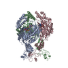
Journal: Proc Natl Acad Sci U S A / Year: 2024 Title: Selective 8-oxo-rG stalling occurs in the catalytic core of polynucleotide phosphorylase (PNPase) during degradation. Authors: Lucas G Miller / Wantae Kim / Shawn Schowe / Kathleen Taylor / Runhua Han / Vashita Jain / Raeyeon Park / Mark Sherman / Janssen Fang / Haydee Ramirez / Andrew Ellington / Phanourios Tamamis ...Authors: Lucas G Miller / Wantae Kim / Shawn Schowe / Kathleen Taylor / Runhua Han / Vashita Jain / Raeyeon Park / Mark Sherman / Janssen Fang / Haydee Ramirez / Andrew Ellington / Phanourios Tamamis / Marino J E Resendiz / Y Jessie Zhang / Lydia Contreras / Abstract: RNA oxidation, predominantly through the accumulation of 8-oxo-7,8-dihydroguanosine (8-oxo-rG), represents an important biomarker for cellular oxidative stress. Polynucleotide phosphorylase (PNPase) ...RNA oxidation, predominantly through the accumulation of 8-oxo-7,8-dihydroguanosine (8-oxo-rG), represents an important biomarker for cellular oxidative stress. Polynucleotide phosphorylase (PNPase) is a 3'-5' exoribonuclease that has been shown to preferentially recognize 8-oxo-rG-containing RNA and protect cells from oxidative stress. However, the impact of 8-oxo-rG on PNPase-mediated RNA degradation has not been studied. Here, we show that the presence of 8-oxo-rG in RNA leads to catalytic stalling of PNPase through in vitro RNA degradation experiments and electrophoretic analysis. We also link this stalling to the active site of the enzyme through resolution of single-particle cryo-EM structures for PNPase in complex with singly or doubly oxidized RNA oligonucleotides. Following identification of Arg399 as a key residue in recognition of both single and sequential 8-oxo-rG nucleotides, we perform follow-up in vitro analysis to confirm the importance of this residue in 8-oxo-rG-specific PNPase stalling. Finally, we investigate the effects of mutations to active site residues implicated in 8-oxo-rG binding through cell growth experiments under HO-induced oxidative stress. Specifically, Arg399 mutations show significant effects on cell growth under oxidative stress. Overall, we demonstrate that 8-oxo-rG-specific stalling of PNPase is relevant to bacterial survival under oxidative stress and speculate that this enzyme might associate with other cellular factors to mediate this stress.
In the structure databanks used in Yorodumi, some data are registered as the other names, "COVID-19 virus" and "2019-nCoV". Here are the details of the virus and the list of structure data.
Jan 31, 2019. EMDB accession codes are about to change! (news from PDBe EMDB page)
EMDB accession codes are about to change! (news from PDBe EMDB page)
The allocation of 4 digits for EMDB accession codes will soon come to an end. Whilst these codes will remain in use, new EMDB accession codes will include an additional digit and will expand incrementally as the available range of codes is exhausted. The current 4-digit format prefixed with “EMD-” (i.e. EMD-XXXX) will advance to a 5-digit format (i.e. EMD-XXXXX), and so on. It is currently estimated that the 4-digit codes will be depleted around Spring 2019, at which point the 5-digit format will come into force.
The EM Navigator/Yorodumi systems omit the EMD- prefix.
Related info.:Q: What is EMD? / ID/Accession-code notation in Yorodumi/EM Navigator
Yorodumi is a browser for structure data from EMDB, PDB, SASBDB, etc.
This page is also the successor to EM Navigator detail page, and also detail information page/front-end page for Omokage search.
The word "yorodu" (or yorozu) is an old Japanese word meaning "ten thousand". "mi" (miru) is to see.
Related info.:EMDB / PDB / SASBDB / Comparison of 3 databanks / Yorodumi Search / Aug 31, 2016. New EM Navigator & Yorodumi / Yorodumi Papers / Jmol/JSmol / Function and homology information / Changes in new EM Navigator and Yorodumi
 Movie
Movie Controller


 Open data
 Basic information
 Map data
 Sample
 Keywords
 Function and homology information
 Authors
 United States, 1 items
  Citation
 Structure visualization
 Downloads & links
 emd_43092.png
 http://ftp.pdbj.org/pub/emdb/structures/EMD-43092
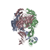


 Z (Sec.)
 Y (Row.)
 X (Col.)


 Sample components
 Processing
 Electron microscopy
 FIELD EMISSION GUN
