 ムービー
ムービー コントローラー


+ データを開く

- 基本情報
基本情報
| 登録情報 |  | |||||||||
|---|---|---|---|---|---|---|---|---|---|---|
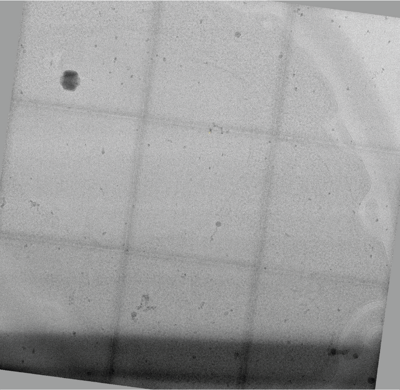
| タイトル | 3x3 tiled montage tomogram of a yeast lamella imaged with a square electron beam | |||||||||
 マップデータ マップデータ | ||||||||||
 試料 試料 |
| |||||||||
 キーワード キーワード | FIB-milled yeast / UNKNOWN FUNCTION | |||||||||
| 生物種 |  | |||||||||
| 手法 | 電子線トモグラフィー法 | |||||||||
 データ登録者 データ登録者 | Chua EYD / Alink LM / Kopylov M / Johnston J / Einsenstein F / de Marco A | |||||||||
| 資金援助 |  米国, 2件 米国, 2件
| |||||||||
 引用 引用 | ジャーナル: Nat Methods / 年: 2024 タイトル: Square beams for optimal tiling in transmission electron microscopy. 著者: Eugene Y D Chua / Lambertus M Alink / Mykhailo Kopylov / Jake D Johnston / Fabian Eisenstein / Alex de Marco /  要旨: Imaging large fields of view at a high magnification requires tiling. Transmission electron microscopes typically have round beam profiles; therefore, tiling across a large area is either imperfect ...Imaging large fields of view at a high magnification requires tiling. Transmission electron microscopes typically have round beam profiles; therefore, tiling across a large area is either imperfect or results in uneven exposures, a problem for dose-sensitive samples. Here, we introduce a square electron beam that can easily be retrofitted in existing microscopes, and demonstrate its application, showing that it can tile nearly perfectly and deliver cryo-electron microscopy imaging with a resolution comparable to conventional set-ups. | |||||||||
| 履歴 |
|
- 構造の表示
構造の表示
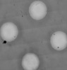
| 添付画像 |
|---|
- ダウンロードとリンク
-EMDBアーカイブ
| マップデータ | emd_42879.map.gz | 12.7 GB |  EMDBマップデータ形式 EMDBマップデータ形式 | |
|---|---|---|---|---|
| ヘッダ (付随情報) | emd-42879-v30.xmlemd-42879.xml | 10.3 KB 10.3 KB | 表示 表示 | EMDBヘッダ |
| 画像 |  emd_42879.png emd_42879.png | 202.8 KB | ||
| Filedesc metadata | emd-42879.cif.gz | 4.1 KB | ||
| アーカイブディレクトリ |  http://ftp.pdbj.org/pub/emdb/structures/EMD-42879ftp://ftp.pdbj.org/pub/emdb/structures/EMD-42879 http://ftp.pdbj.org/pub/emdb/structures/EMD-42879ftp://ftp.pdbj.org/pub/emdb/structures/EMD-42879 | HTTPS FTP |
-検証レポート
| 文書・要旨 | emd_42879_validation.pdf.gz | 700.1 KB | 表示 | EMDB検証レポート |
|---|---|---|---|---|
| 文書・詳細版 | emd_42879_full_validation.pdf.gz | 699.6 KB | 表示 | |
| XML形式データ | emd_42879_validation.xml.gz | 4.8 KB | 表示 | |
| CIF形式データ | emd_42879_validation.cif.gz | 5.3 KB | 表示 | |
| アーカイブディレクトリ | https://ftp.pdbj.org/pub/emdb/validation_reports/EMD-42879ftp://ftp.pdbj.org/pub/emdb/validation_reports/EMD-42879 | HTTPS FTP |
-関連構造データ


-リンク
| EMDBのページ | EMDB (EBI/PDBe) / EMDataResource |
|---|
-マップ
| ファイル | ダウンロード / ファイル: emd_42879.map.gz / 形式: CCP4 / 大きさ: 13.7 GB / タイプ: IMAGE STORED AS FLOATING POINT NUMBER (4 BYTES) | ||||||||||||||||||||||||||||||||
|---|---|---|---|---|---|---|---|---|---|---|---|---|---|---|---|---|---|---|---|---|---|---|---|---|---|---|---|---|---|---|---|---|---|
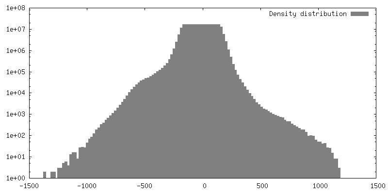
| 投影像・断面図 | 画像のコントロール
画像は Spider により作成 これらの図は立方格子座標系で作成されたものです | ||||||||||||||||||||||||||||||||
| ボクセルのサイズ | X=Y=Z: 8 Å | ||||||||||||||||||||||||||||||||
| 密度 |
| ||||||||||||||||||||||||||||||||
| 対称性 | 空間群: 1 | ||||||||||||||||||||||||||||||||
| 詳細 | EMDB XML:
|
 Z (Sec.)
 Y (Row.)
 X (Col.)


-添付データ
- 試料の構成要素
-全体 : Yeast
| 全体 | 名称: |
|---|---|
| 要素 |
|
-超分子 #1: Yeast
| 超分子 | 名称: Yeast / タイプ: cell / ID: 1 / 親要素: 0 |
|---|---|
| 由来(天然) | 生物種: |
-実験情報
-構造解析
 解析 解析 | 電子線トモグラフィー法 |
|---|---|
| 試料の集合状態 | cell |
-試料調製
| 緩衝液 | pH: 7.5 / 詳細: YPD media |
|---|---|
| グリッド | モデル: Quantifoil R2/2 / 材質: COPPER / メッシュ: 200 / 支持フィルム - 材質: CARBON / 支持フィルム - トポロジー: HOLEY / 前処理 - タイプ: GLOW DISCHARGE / 前処理 - 雰囲気: OTHER / 詳細: Gatan Solarus I |
| 詳細 | Extrapolated OD 600 ~20 |
| 加圧凍結法 | 装置: OTHER 詳細: Sample was high-pressure frozen directly on a 200 mesh grid sandwiched between two flat sides of 3 mm planchettes. The value given for _em_high_pressure_freezing.instrument is Wohlwend HPF ...詳細: Sample was high-pressure frozen directly on a 200 mesh grid sandwiched between two flat sides of 3 mm planchettes. The value given for _em_high_pressure_freezing.instrument is Wohlwend HPF Compact 01. This is not in a list of allowed values {'LEICA EM HPM100', 'OTHER', 'LEICA EM PACT', 'BAL-TEC HPM 010', 'EMS-002 RAPID IMMERSION FREEZER', 'LEICA EM PACT2'} so OTHER is written into the XML file. |
| Cryo protectant | 5% glycerol |
| 切片作成 | 集束イオンビーム - 装置: OTHER / 集束イオンビーム - イオン: OTHER / 集束イオンビーム - 電圧: 30 / 集束イオンビーム - 電流: 2 / 集束イオンビーム - 時間: 300 / 集束イオンビーム - 温度: 77 K / 集束イオンビーム - Initial thickness: 500 / 集束イオンビーム - 最終 厚さ: 200 集束イオンビーム - 詳細: Lamellae were prepared using TFS AutoTEM. The settings specified are for the final polishing only. The complete set of parameters is described in the Waffle Method ...集束イオンビーム - 詳細: Lamellae were prepared using TFS AutoTEM. The settings specified are for the final polishing only. The complete set of parameters is described in the Waffle Method protocol paper here: https://ncbi.nlm.nih.gov/pmc/articles/PMC9795037/. The value given for _em_focused_ion_beam.instrument is Aquilos 2. This is not in a list of allowed values {'DB235', 'OTHER'} so OTHER is written into the XML file. |
- 電子顕微鏡法
| 顕微鏡 | FEI TITAN KRIOS |
|---|---|
| 撮影 | フィルム・検出器のモデル: GATAN K3 BIOQUANTUM (6k x 4k) 平均電子線量: 2.55 e/Å2 |
| 電子線 | 加速電圧: 300 kV / 電子線源:  FIELD EMISSION GUN FIELD EMISSION GUN |
| 電子光学系 | C2レンズ絞り径: 50.0 µm / 照射モード: FLOOD BEAM / 撮影モード: BRIGHT FIELD / Cs: 2.7 mm / 最大 デフォーカス(公称値): 5.0 µm / 最小 デフォーカス(公称値): 2.5 µm |
| 実験機器 |  モデル: Titan Krios / 画像提供: FEI Company |
-画像解析
| 最終 再構成 | ソフトウェア - 名称: TOMO3D / 使用した粒子像数: 279 |
|---|
