 Movie
Movie Controller

[English] 日本語
 Yorodumi
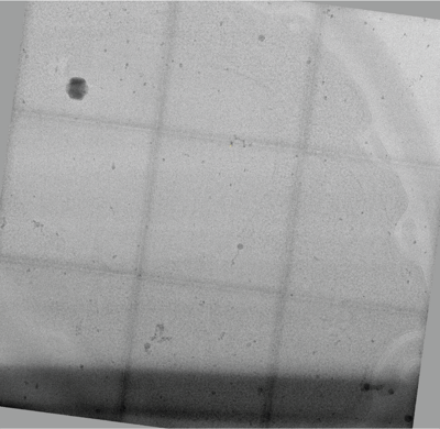
- EMDB-42879: 3x3 tiled montage tomogram of a yeast lamella imaged with a squar... -
+ Open data

- Basic information
| Entry |  | |||||||||
|---|---|---|---|---|---|---|---|---|---|---|
| Title | 3x3 tiled montage tomogram of a yeast lamella imaged with a square electron beam | |||||||||
 Map data Map data | ||||||||||
 Sample Sample |
| |||||||||
 Keywords Keywords | FIB-milled yeast / UNKNOWN FUNCTION | |||||||||
| Biological species |  | |||||||||
| Method | electron tomography | |||||||||
 Authors Authors | Chua EYD / Alink LM / Kopylov M / Johnston J / Einsenstein F / de Marco A | |||||||||
| Funding support |  United States, 2 items United States, 2 items
| |||||||||
 Citation Citation | Journal: Nat Methods / Year: 2024 Title: Square beams for optimal tiling in transmission electron microscopy. Authors: Eugene Y D Chua / Lambertus M Alink / Mykhailo Kopylov / Jake D Johnston / Fabian Eisenstein / Alex de Marco /  Abstract: Imaging large fields of view at a high magnification requires tiling. Transmission electron microscopes typically have round beam profiles; therefore, tiling across a large area is either imperfect ...Imaging large fields of view at a high magnification requires tiling. Transmission electron microscopes typically have round beam profiles; therefore, tiling across a large area is either imperfect or results in uneven exposures, a problem for dose-sensitive samples. Here, we introduce a square electron beam that can easily be retrofitted in existing microscopes, and demonstrate its application, showing that it can tile nearly perfectly and deliver cryo-electron microscopy imaging with a resolution comparable to conventional set-ups. | |||||||||
| History |
|
- Structure visualization
| Supplemental images |
|---|
- Downloads & links
-EMDB archive
| Map data | emd_42879.map.gz | 12.7 GB |  EMDB map data format EMDB map data format | |
|---|---|---|---|---|
| Header (meta data) | emd-42879-v30.xmlemd-42879.xml | 10.3 KB 10.3 KB | Display Display | EMDB header |
| Images |  emd_42879.png emd_42879.png | 202.8 KB | ||
| Filedesc metadata | emd-42879.cif.gz | 4.1 KB | ||
| Archive directory |  http://ftp.pdbj.org/pub/emdb/structures/EMD-42879ftp://ftp.pdbj.org/pub/emdb/structures/EMD-42879 http://ftp.pdbj.org/pub/emdb/structures/EMD-42879ftp://ftp.pdbj.org/pub/emdb/structures/EMD-42879 | HTTPS FTP |
-Validation report
| Summary document | emd_42879_validation.pdf.gz | 700.1 KB | Display | EMDB validaton report |
|---|---|---|---|---|
| Full document | emd_42879_full_validation.pdf.gz | 699.6 KB | Display | |
| Data in XML | emd_42879_validation.xml.gz | 4.8 KB | Display | |
| Data in CIF | emd_42879_validation.cif.gz | 5.3 KB | Display | |
| Arichive directory | https://ftp.pdbj.org/pub/emdb/validation_reports/EMD-42879ftp://ftp.pdbj.org/pub/emdb/validation_reports/EMD-42879 | HTTPS FTP |
-Related structure data


-Links
| EMDB pages | EMDB (EBI/PDBe) / EMDataResource |
|---|
-Map
| File | Download / File: emd_42879.map.gz / Format: CCP4 / Size: 13.7 GB / Type: IMAGE STORED AS FLOATING POINT NUMBER (4 BYTES) | ||||||||||||||||||||||||||||||||
|---|---|---|---|---|---|---|---|---|---|---|---|---|---|---|---|---|---|---|---|---|---|---|---|---|---|---|---|---|---|---|---|---|---|
| Projections & slices | Image control
Images are generated by Spider. generated in cubic-lattice coordinate | ||||||||||||||||||||||||||||||||
| Voxel size | X=Y=Z: 8 Å | ||||||||||||||||||||||||||||||||
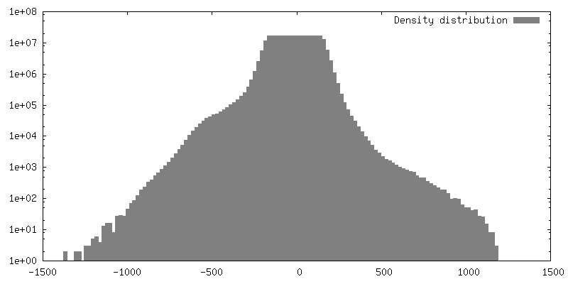
| Density |
| ||||||||||||||||||||||||||||||||
| Symmetry | Space group: 1 | ||||||||||||||||||||||||||||||||
| Details | EMDB XML:
|
 Z (Sec.)
 Y (Row.)
 X (Col.)


-Supplemental data
- Sample components
-Entire : Yeast
| Entire | Name: |
|---|---|
| Components |
|
-Supramolecule #1: Yeast
| Supramolecule | Name: Yeast / type: cell / ID: 1 / Parent: 0 |
|---|---|
| Source (natural) | Organism: |
-Experimental details
-Structure determination
 Processing Processing | electron tomography |
|---|---|
| Aggregation state | cell |
-Sample preparation
| Buffer | pH: 7.5 / Details: YPD media |
|---|---|
| Grid | Model: Quantifoil R2/2 / Material: COPPER / Mesh: 200 / Support film - Material: CARBON / Support film - topology: HOLEY / Pretreatment - Type: GLOW DISCHARGE / Pretreatment - Atmosphere: OTHER / Details: Gatan Solarus I |
| Details | Extrapolated OD 600 ~20 |
| High pressure freezing | Instrument: OTHER Details: Sample was high-pressure frozen directly on a 200 mesh grid sandwiched between two flat sides of 3 mm planchettes. The value given for _em_high_pressure_freezing.instrument is Wohlwend HPF ...Details: Sample was high-pressure frozen directly on a 200 mesh grid sandwiched between two flat sides of 3 mm planchettes. The value given for _em_high_pressure_freezing.instrument is Wohlwend HPF Compact 01. This is not in a list of allowed values {'LEICA EM HPM100', 'OTHER', 'LEICA EM PACT', 'BAL-TEC HPM 010', 'EMS-002 RAPID IMMERSION FREEZER', 'LEICA EM PACT2'} so OTHER is written into the XML file. |
| Cryo protectant | 5% glycerol |
| Sectioning | Focused ion beam - Instrument: OTHER / Focused ion beam - Ion: OTHER / Focused ion beam - Voltage: 30 / Focused ion beam - Current: 2 / Focused ion beam - Duration: 300 / Focused ion beam - Temperature: 77 K / Focused ion beam - Initial thickness: 500 / Focused ion beam - Final thickness: 200 Focused ion beam - Details: Lamellae were prepared using TFS AutoTEM. The settings specified are for the final polishing only. The complete set of parameters is described in the Waffle Method ...Focused ion beam - Details: Lamellae were prepared using TFS AutoTEM. The settings specified are for the final polishing only. The complete set of parameters is described in the Waffle Method protocol paper here: https://ncbi.nlm.nih.gov/pmc/articles/PMC9795037/. The value given for _em_focused_ion_beam.instrument is Aquilos 2. This is not in a list of allowed values {'DB235', 'OTHER'} so OTHER is written into the XML file. |
- Electron microscopy
| Microscope | FEI TITAN KRIOS |
|---|---|
| Image recording | Film or detector model: GATAN K3 BIOQUANTUM (6k x 4k) / Average electron dose: 2.55 e/Å2 |
| Electron beam | Acceleration voltage: 300 kV / Electron source:  FIELD EMISSION GUN FIELD EMISSION GUN |
| Electron optics | C2 aperture diameter: 50.0 µm / Illumination mode: FLOOD BEAM / Imaging mode: BRIGHT FIELD / Cs: 2.7 mm / Nominal defocus max: 5.0 µm / Nominal defocus min: 2.5 µm |
| Experimental equipment |  Model: Titan Krios / Image courtesy: FEI Company |
-Image processing
| Final reconstruction | Software - Name: TOMO3D / Number images used: 279 |
|---|
