 Movie
Movie Controller


+ Open data

- Basic information
| Entry |  | |||||||||
|---|---|---|---|---|---|---|---|---|---|---|
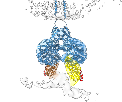
| Title | ACE2 dimer bound to two RBD in membrane | |||||||||
 Map data Map data | ||||||||||
 Sample Sample |
| |||||||||
 Keywords Keywords | Complex / VIRAL PROTEIN / Cell Receptor | |||||||||
| Biological species |   Severe acute respiratory syndrome coronavirus 2 Severe acute respiratory syndrome coronavirus 2 | |||||||||
| Method | subtomogram averaging / cryo EM / Resolution: 18.0 Å | |||||||||
 Authors Authors | Qin Z / Li W / Grunst MW / Mothes W | |||||||||
| Funding support |  United States, 1 items United States, 1 items
| |||||||||
 Citation Citation | Journal: Science / Year: 2024 Title: Structure and inhibition of SARS-CoV-2 spike refolding in membranes. Authors: Michael W Grunst / Zhuan Qin / Esteban Dodero-Rojas / Shilei Ding / Jérémie Prévost / Yaozong Chen / Yanping Hu / Marzena Pazgier / Shenping Wu / Xuping Xie / Andrés Finzi / José N ...Authors: Michael W Grunst / Zhuan Qin / Esteban Dodero-Rojas / Shilei Ding / Jérémie Prévost / Yaozong Chen / Yanping Hu / Marzena Pazgier / Shenping Wu / Xuping Xie / Andrés Finzi / José N Onuchic / Paul C Whitford / Walther Mothes / Wenwei Li /  Abstract: The severe acute respiratory syndrome coronavirus 2 (SARS-CoV-2) spike protein binds the receptor angiotensin converting enzyme 2 (ACE2) and drives virus-host membrane fusion through refolding of its ...The severe acute respiratory syndrome coronavirus 2 (SARS-CoV-2) spike protein binds the receptor angiotensin converting enzyme 2 (ACE2) and drives virus-host membrane fusion through refolding of its S2 domain. Whereas the S1 domain contains high sequence variability, the S2 domain is conserved and is a promising pan-betacoronavirus vaccine target. We applied cryo-electron tomography to capture intermediates of S2 refolding and understand inhibition by antibodies to the S2 stem-helix. Subtomogram averaging revealed ACE2 dimers cross-linking spikes before transitioning into S2 intermediates, which were captured at various stages of refolding. Pan-betacoronavirus neutralizing antibodies targeting the S2 stem-helix bound to and inhibited refolding of spike prehairpin intermediates. Combined with molecular dynamics simulations, these structures elucidate the process of SARS-CoV-2 entry and reveal how pan-betacoronavirus S2-targeting antibodies neutralize infectivity by arresting prehairpin intermediates. | |||||||||
| History |
|
- Structure visualization
| Supplemental images |
|---|
- Downloads & links
-EMDB archive
| Map data | emd_42876.map.gz | 1.8 MB |  EMDB map data format EMDB map data format | |
|---|---|---|---|---|
| Header (meta data) | emd-42876-v30.xmlemd-42876.xml | 14 KB 14 KB | Display Display | EMDB header |
| FSC (resolution estimation) | emd_42876_fsc.xml | 3 KB | Display | FSC data file |
| Images |  emd_42876.png emd_42876.png | 86.5 KB | ||
| Filedesc metadata | emd-42876.cif.gz | 4.1 KB | ||
| Others | emd_42876_half_map_1.map.gzemd_42876_half_map_2.map.gz | 1.8 MB 1.8 MB | ||
| Archive directory |  http://ftp.pdbj.org/pub/emdb/structures/EMD-42876ftp://ftp.pdbj.org/pub/emdb/structures/EMD-42876 http://ftp.pdbj.org/pub/emdb/structures/EMD-42876ftp://ftp.pdbj.org/pub/emdb/structures/EMD-42876 | HTTPS FTP |
-Related structure data


-Links
| EMDB pages | EMDB (EBI/PDBe) / EMDataResource |
|---|
-Map
| File | Download / File: emd_42876.map.gz / Format: CCP4 / Size: 2 MB / Type: IMAGE STORED AS FLOATING POINT NUMBER (4 BYTES) | ||||||||||||||||||||||||||||||||||||
|---|---|---|---|---|---|---|---|---|---|---|---|---|---|---|---|---|---|---|---|---|---|---|---|---|---|---|---|---|---|---|---|---|---|---|---|---|---|
| Projections & slices | Image control
Images are generated by Spider. | ||||||||||||||||||||||||||||||||||||
| Voxel size | X=Y=Z: 2.692 Å | ||||||||||||||||||||||||||||||||||||
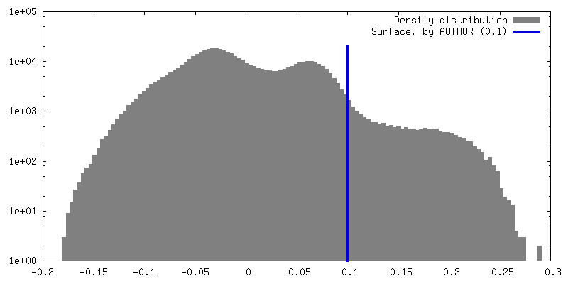
| Density |
| ||||||||||||||||||||||||||||||||||||
| Symmetry | Space group: 1 | ||||||||||||||||||||||||||||||||||||
| Details | EMDB XML:
|
 Z (Sec.)
 Y (Row.)
 X (Col.)


-Supplemental data
- Sample components
-Entire : ACE2 dimer bound to two RBD in membrane
| Entire | Name: ACE2 dimer bound to two RBD in membrane |
|---|---|
| Components |
|
-Supramolecule #1: ACE2 dimer bound to two RBD in membrane
| Supramolecule | Name: ACE2 dimer bound to two RBD in membrane / type: complex / ID: 1 / Parent: 0 / Macromolecule list: #1-#2 |
|---|---|
| Source (natural) | Organism: Severe acute respiratory syndrome coronavirus 2 |
-Experimental details
-Structure determination
| Method | cryo EM |
|---|---|
 Processing Processing | subtomogram averaging |
| Aggregation state | particle |
-Sample preparation
| Buffer | pH: 7.4 |
|---|---|
| Vitrification | Cryogen name: ETHANE |
- Electron microscopy
| Microscope | FEI TITAN KRIOS |
|---|---|
| Specialist optics | Phase plate: VOLTA PHASE PLATE / Energy filter - Name: GIF Quantum LS / Energy filter - Slit width: 20 eV |
| Image recording | Film or detector model: GATAN K3 BIOQUANTUM (6k x 4k) / Average electron dose: 3.0 e/Å2 |
| Electron beam | Acceleration voltage: 300 kV / Electron source:  FIELD EMISSION GUN FIELD EMISSION GUN |
| Electron optics | C2 aperture diameter: 50.0 µm / Calibrated defocus max: 0.8 µm / Calibrated defocus min: 0.2 µm / Illumination mode: OTHER / Imaging mode: BRIGHT FIELD / Cs: 2.7 mm / Nominal defocus max: 0.5 µm / Nominal defocus min: 0.5 µm / Nominal magnification: 64000 |
| Sample stage | Specimen holder model: FEI TITAN KRIOS AUTOGRID HOLDER / Cooling holder cryogen: NITROGEN |
| Experimental equipment |  Model: Titan Krios / Image courtesy: FEI Company |
-Image processing
| Final reconstruction | Applied symmetry - Point group: C1 (asymmetric) / Resolution.type: BY AUTHOR / Resolution: 18.0 Å / Resolution method: FSC 0.5 CUT-OFF / Software - Name: I3 / Number subtomograms used: 2279 |
|---|---|
| Extraction | Number tomograms: 141 / Number images used: 5916 |
| Final angle assignment | Type: NOT APPLICABLE |
| FSC plot (resolution estimation) | 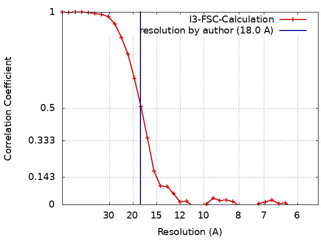 |
