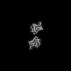
Journal: Sci Adv / Year: 2024 Title: Mouse α-synuclein fibrils are structurally and functionally distinct from human fibrils associated with Lewy body diseases. Authors: Arpine Sokratian / Ye Zhou / Meltem Tatli / Kevin J Burbidge / Enquan Xu / Elizabeth Viverette / Sonia Donzelli / Addison M Duda / Yuan Yuan / Huizhong Li / Samuel Strader / Nirali Patel / ...Authors: Arpine Sokratian / Ye Zhou / Meltem Tatli / Kevin J Burbidge / Enquan Xu / Elizabeth Viverette / Sonia Donzelli / Addison M Duda / Yuan Yuan / Huizhong Li / Samuel Strader / Nirali Patel / Lauren Shiell / Tuyana Malankhanova / Olivia Chen / Joseph R Mazzulli / Lalith Perera / Henning Stahlberg / Mario Borgnia / Alberto Bartesaghi / Hilal A Lashuel / Andrew B West / Abstract: The intricate process of α-synuclein aggregation and fibrillization holds pivotal roles in Parkinson's disease (PD) and multiple system atrophy (MSA). While mouse α-synuclein can fibrillize in ...The intricate process of α-synuclein aggregation and fibrillization holds pivotal roles in Parkinson's disease (PD) and multiple system atrophy (MSA). While mouse α-synuclein can fibrillize in vitro, whether these fibrils commonly used in research to induce this process or form can reproduce structures in the human brain remains unknown. Here, we report the first atomic structure of mouse α-synuclein fibrils, which was solved in parallel by two independent teams. The structure shows striking similarity to MSA-amplified and PD-associated E46K fibrils. However, mouse α-synuclein fibrils display altered packing arrangements, reduced hydrophobicity, and heightened fragmentation sensitivity and evoke only weak immunological responses. Furthermore, mouse α-synuclein fibrils exhibit exacerbated pathological spread in neurons and humanized α-synuclein mice. These findings provide critical insights into the structural underpinnings of α-synuclein pathogenicity and emphasize a need to reassess the role of mouse α-synuclein fibrils in the development of related diagnostic probes and therapeutic interventions.
In the structure databanks used in Yorodumi, some data are registered as the other names, "COVID-19 virus" and "2019-nCoV". Here are the details of the virus and the list of structure data.
Jan 31, 2019. EMDB accession codes are about to change! (news from PDBe EMDB page)
EMDB accession codes are about to change! (news from PDBe EMDB page)
The allocation of 4 digits for EMDB accession codes will soon come to an end. Whilst these codes will remain in use, new EMDB accession codes will include an additional digit and will expand incrementally as the available range of codes is exhausted. The current 4-digit format prefixed with “EMD-” (i.e. EMD-XXXX) will advance to a 5-digit format (i.e. EMD-XXXXX), and so on. It is currently estimated that the 4-digit codes will be depleted around Spring 2019, at which point the 5-digit format will come into force.
The EM Navigator/Yorodumi systems omit the EMD- prefix.
Related info.:Q: What is EMD? / ID/Accession-code notation in Yorodumi/EM Navigator
Yorodumi is a browser for structure data from EMDB, PDB, SASBDB, etc.
This page is also the successor to EM Navigator detail page, and also detail information page/front-end page for Omokage search.
The word "yorodu" (or yorozu) is an old Japanese word meaning "ten thousand". "mi" (miru) is to see.
Related info.:EMDB / PDB / SASBDB / Comparison of 3 databanks / Yorodumi Search / Aug 31, 2016. New EM Navigator & Yorodumi / Yorodumi Papers / Jmol/JSmol / Function and homology information / Changes in new EM Navigator and Yorodumi
 Movie
Movie Controller

 Yorodumi
 Open data
 Basic information
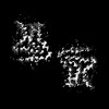
 Map data
 Sample
 Keywords
 Function and homology information
 Authors
 United States, 1 items
  Citation
 Structure visualization
 Downloads & links
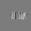
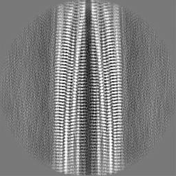
 emd_42294.png
 http://ftp.pdbj.org/pub/emdb/structures/EMD-42294

 Z (Sec.)
 Y (Row.)
 X (Col.)


 Sample components
 Processing
 Electron microscopy
 FIELD EMISSION GUN

