 Movie
Movie Controller

[English] 日本語
 Yorodumi
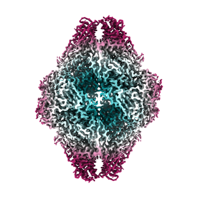
- EMDB-41919: Structure of E.coli beta-Galactosidase from a multi-species dataset -
+ Open data

- Basic information
| Entry |  | |||||||||
|---|---|---|---|---|---|---|---|---|---|---|
| Title | Structure of E.coli beta-Galactosidase from a multi-species dataset | |||||||||
 Map data Map data | Final map sharp | |||||||||
 Sample Sample |
| |||||||||
 Keywords Keywords | hydrolase / D2 / beta-gal | |||||||||
| Biological species |  | |||||||||
| Method | single particle reconstruction / cryo EM / Resolution: 2.73 Å | |||||||||
 Authors Authors | Kopylov M / Bobe D / Eng ET | |||||||||
| Funding support |  United States, 1 items United States, 1 items
| |||||||||
 Citation Citation | Journal: Acta Crystallogr F Struct Biol Commun / Year: 2024 Title: Multi-species cryoEM calibration and workflow verification standard. Authors: Daija Bobe / Mykhailo Kopylov / Jessalyn Miller / Aaron P Owji / Edward T Eng / Abstract: Cryogenic electron microscopy (cryoEM) is a rapidly growing structural biology modality that has been successful in revealing molecular details of biological systems. However, unlike established ...Cryogenic electron microscopy (cryoEM) is a rapidly growing structural biology modality that has been successful in revealing molecular details of biological systems. However, unlike established biophysical and analytical techniques with calibration standards, cryoEM has lacked comprehensive biological test samples. Here, a cryoEM calibration sample consisting of a mixture of compatible macromolecules is introduced that can not only be used for resolution optimization, but also provides multiple reference points for evaluating instrument performance, data quality and image-processing workflows in a single experiment. This combined test specimen provides researchers with a reference point for validating their cryoEM pipeline, benchmarking their methodologies and testing new algorithms. | |||||||||
| History |
|
- Structure visualization
| Supplemental images |
|---|
- Downloads & links
-EMDB archive
| Map data | emd_41919.map.gz | 167.9 MB |  EMDB map data format EMDB map data format | |
|---|---|---|---|---|
| Header (meta data) | emd-41919-v30.xmlemd-41919.xml | 15.7 KB 15.7 KB | Display Display | EMDB header |
| FSC (resolution estimation) | emd_41919_fsc.xml | 11.8 KB | Display | FSC data file |
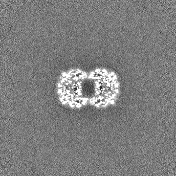
| Images |  emd_41919.png emd_41919.png | 143.4 KB | ||
| Masks | emd_41919_msk_1.map | 178 MB | Mask map | |
| Filedesc metadata | emd-41919.cif.gz | 4.3 KB | ||
| Others | emd_41919_half_map_1.map.gzemd_41919_half_map_2.map.gz | 165 MB 165 MB | ||
| Archive directory |  http://ftp.pdbj.org/pub/emdb/structures/EMD-41919ftp://ftp.pdbj.org/pub/emdb/structures/EMD-41919 http://ftp.pdbj.org/pub/emdb/structures/EMD-41919ftp://ftp.pdbj.org/pub/emdb/structures/EMD-41919 | HTTPS FTP |
-Related structure data


-Links
| EMDB pages | EMDB (EBI/PDBe) / EMDataResource |
|---|
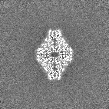
-Map
| File | Download / File: emd_41919.map.gz / Format: CCP4 / Size: 178 MB / Type: IMAGE STORED AS FLOATING POINT NUMBER (4 BYTES) | ||||||||||||||||||||||||||||||||||||
|---|---|---|---|---|---|---|---|---|---|---|---|---|---|---|---|---|---|---|---|---|---|---|---|---|---|---|---|---|---|---|---|---|---|---|---|---|---|
| Annotation | Final map sharp | ||||||||||||||||||||||||||||||||||||
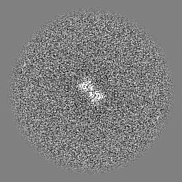
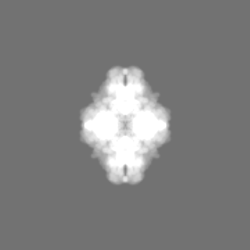
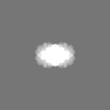
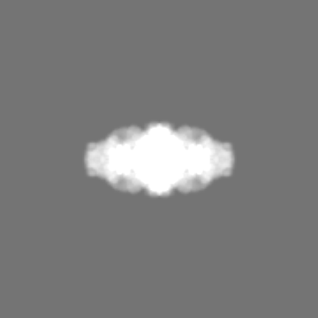
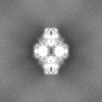
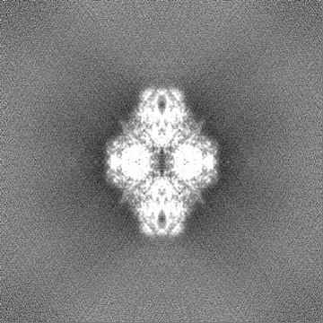
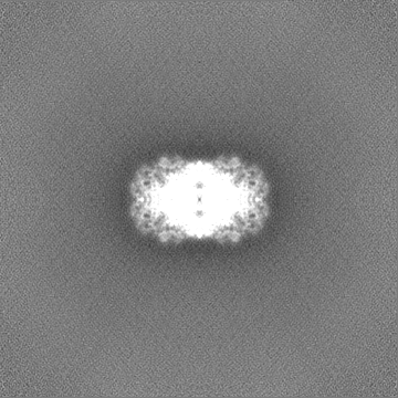
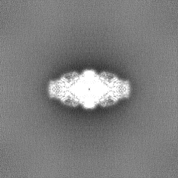
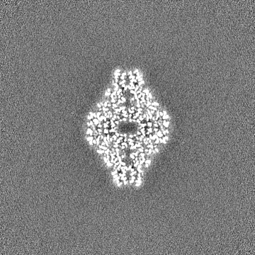
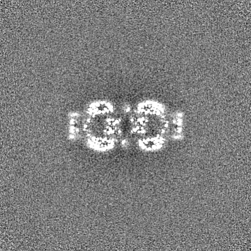
| Projections & slices | Image control
Images are generated by Spider. | ||||||||||||||||||||||||||||||||||||
| Voxel size | X=Y=Z: 1.096 Å | ||||||||||||||||||||||||||||||||||||
| Density |
| ||||||||||||||||||||||||||||||||||||
| Symmetry | Space group: 1 | ||||||||||||||||||||||||||||||||||||
| Details | EMDB XML:
|
 Z (Sec.)
 Y (Row.)
 X (Col.)


-Supplemental data
-Mask #1
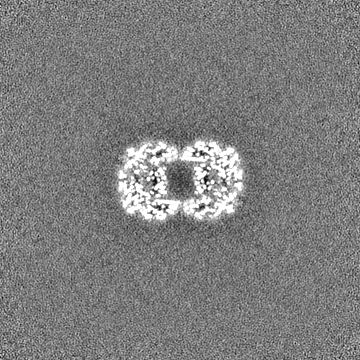
| File | emd_41919_msk_1.map | ||||||||||||
|---|---|---|---|---|---|---|---|---|---|---|---|---|---|
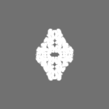
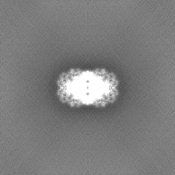
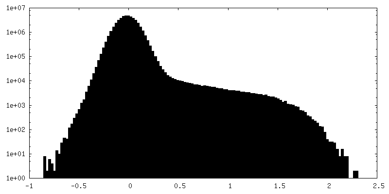
| Projections & Slices |
| ||||||||||||
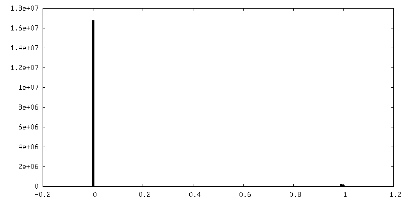
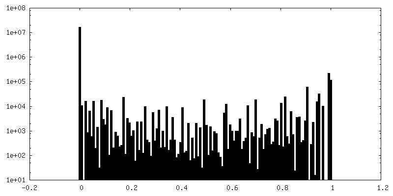
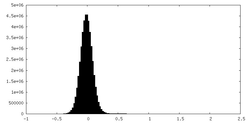
| Density Histograms |

-Half map: Halfmap A
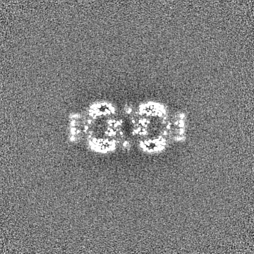
| File | emd_41919_half_map_1.map | ||||||||||||
|---|---|---|---|---|---|---|---|---|---|---|---|---|---|
| Annotation | Halfmap A | ||||||||||||
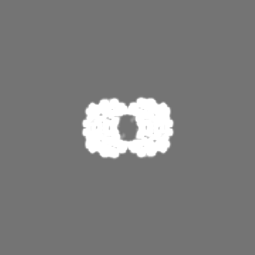
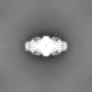
| Projections & Slices |
| ||||||||||||
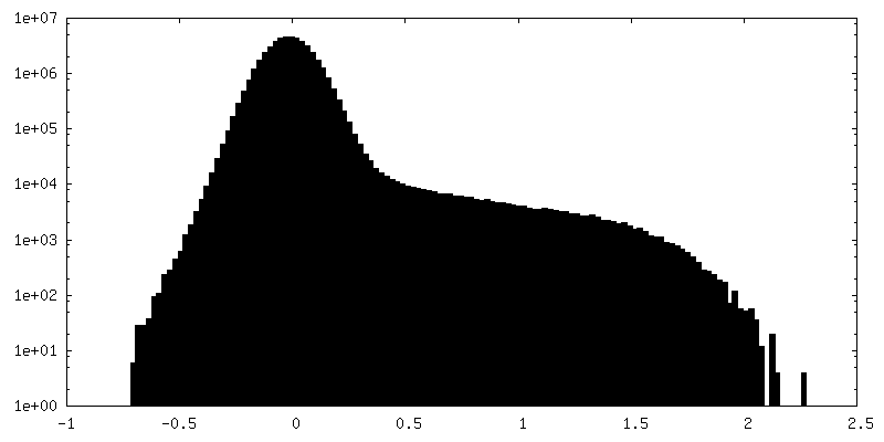
| Density Histograms |

-Half map: Halfmap B
| File | emd_41919_half_map_2.map | ||||||||||||
|---|---|---|---|---|---|---|---|---|---|---|---|---|---|
| Annotation | Halfmap B | ||||||||||||
| Projections & Slices |
| ||||||||||||
| Density Histograms |
- Sample components
-Entire : beta-Galactosidase from Escherichia coli in complex with 2-phenyl...
| Entire | Name: beta-Galactosidase from Escherichia coli in complex with 2-phenylethyl beta-D-thiogalactoside |
|---|---|
| Components |
|
-Supramolecule #1: beta-Galactosidase from Escherichia coli in complex with 2-phenyl...
| Supramolecule | Name: beta-Galactosidase from Escherichia coli in complex with 2-phenylethyl beta-D-thiogalactoside type: complex / ID: 1 / Parent: 0 |
|---|---|
| Source (natural) | Organism: |
| Molecular weight | Theoretical: 2.5 MDa |
-Experimental details
-Structure determination
| Method | cryo EM |
|---|---|
 Processing Processing | single particle reconstruction |
| Aggregation state | particle |
-Sample preparation
| Concentration | 1 mg/mL |
|---|---|
| Buffer | pH: 7.5 |
| Vitrification | Cryogen name: ETHANE |
- Electron microscopy
| Microscope | TFS KRIOS |
|---|---|
| Image recording | Film or detector model: GATAN K2 SUMMIT (4k x 4k) / Average electron dose: 70.0 e/Å2 |
| Electron beam | Acceleration voltage: 300 kV / Electron source:  FIELD EMISSION GUN FIELD EMISSION GUN |
| Electron optics | Illumination mode: FLOOD BEAM / Imaging mode: BRIGHT FIELD / Nominal defocus max: 2.5 µm / Nominal defocus min: 1.5 µm |
| Experimental equipment |  Model: Titan Krios / Image courtesy: FEI Company |

