 ムービー
ムービー コントローラー


+ データを開く

- 基本情報
基本情報
| 登録情報 |  | |||||||||
|---|---|---|---|---|---|---|---|---|---|---|
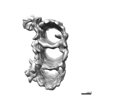
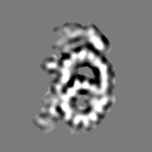
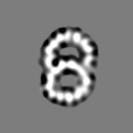
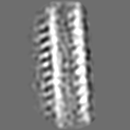
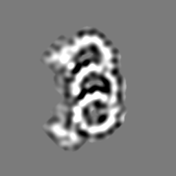
| タイトル | Subtomogram Averaging of the Triplet from Mouse Centrioles | |||||||||
 マップデータ マップデータ | Subtomogram averaging of complete triplet from proximal centriole | |||||||||
 試料 試料 |
| |||||||||
 キーワード キーワード | Inhibitor / SIGNALING PROTEIN | |||||||||
| 生物種 |  | |||||||||
| 手法 | サブトモグラム平均法 / クライオ電子顕微鏡法 / 解像度: 40.0 Å | |||||||||
 データ登録者 データ登録者 | Zhang Z / Moye A / He F / Chen M / Agosto AA / Wensel TG | |||||||||
| 資金援助 |  米国, 1件 米国, 1件
| |||||||||
 引用 引用 | ジャーナル: Life Sci Alliance / 年: 2024 タイトル: Centriole and transition zone structures in photoreceptor cilia revealed by cryo-electron tomography. 著者: Zhixian Zhang / Abigail R Moye / Feng He / Muyuan Chen / Melina A Agosto / Theodore G Wensel /   要旨: Primary cilia mediate sensory signaling in multiple organisms and cell types but have structures adapted for specific roles. Structural defects in them lead to devastating diseases known as ...Primary cilia mediate sensory signaling in multiple organisms and cell types but have structures adapted for specific roles. Structural defects in them lead to devastating diseases known as ciliopathies in humans. Key to their functions are structures at their base: the basal body, the transition zone, the "Y-shaped links," and the "ciliary necklace." We have used cryo-electron tomography with subtomogram averaging and conventional transmission electron microscopy to elucidate the structures associated with the basal region of the "connecting cilia" of rod outer segments in mouse retina. The longitudinal variations in microtubule (MT) structures and the lumenal scaffold complexes connecting them have been determined, as well as membrane-associated transition zone structures: Y-shaped links connecting MT to the membrane, and ciliary beads connected to them that protrude from the cell surface and form a necklace-like structure. These results represent a clearer structural scaffold onto which molecules identified by genetics, proteomics, and superresolution fluorescence can be placed in our emerging model of photoreceptor sensory cilia. | |||||||||
| 履歴 |
|
- 構造の表示
構造の表示
| 添付画像 |
|---|
- ダウンロードとリンク
-EMDBアーカイブ
| マップデータ | emd_41812.map.gz | 49.6 MB |  EMDBマップデータ形式 EMDBマップデータ形式 | |
|---|---|---|---|---|
| ヘッダ (付随情報) | emd-41812-v30.xmlemd-41812.xml | 18.6 KB 18.6 KB | 表示 表示 | EMDBヘッダ |
| FSC (解像度算出) | emd_41812_fsc.xml | 9.2 KB | 表示 | FSCデータファイル |
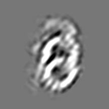
| 画像 |  emd_41812.png emd_41812.png | 42.2 KB | ||
| Filedesc metadata | emd-41812.cif.gz | 4.4 KB | ||
| その他 | emd_41812_additional_1.map.gzemd_41812_additional_2.map.gzemd_41812_additional_3.map.gzemd_41812_half_map_1.map.gzemd_41812_half_map_2.map.gz | 30.9 MB 16.3 MB 16.6 MB 38.7 MB 38.6 MB | ||
| アーカイブディレクトリ |  http://ftp.pdbj.org/pub/emdb/structures/EMD-41812ftp://ftp.pdbj.org/pub/emdb/structures/EMD-41812 http://ftp.pdbj.org/pub/emdb/structures/EMD-41812ftp://ftp.pdbj.org/pub/emdb/structures/EMD-41812 | HTTPS FTP |
-検証レポート
| 文書・要旨 | emd_41812_validation.pdf.gz | 606.5 KB | 表示 | EMDB検証レポート |
|---|---|---|---|---|
| 文書・詳細版 | emd_41812_full_validation.pdf.gz | 606.1 KB | 表示 | |
| XML形式データ | emd_41812_validation.xml.gz | 13.9 KB | 表示 | |
| CIF形式データ | emd_41812_validation.cif.gz | 21.9 KB | 表示 | |
| アーカイブディレクトリ | https://ftp.pdbj.org/pub/emdb/validation_reports/EMD-41812ftp://ftp.pdbj.org/pub/emdb/validation_reports/EMD-41812 | HTTPS FTP |
-関連構造データ


-リンク
| EMDBのページ | EMDB (EBI/PDBe) / EMDataResource |
|---|
-マップ
| ファイル | ダウンロード / ファイル: emd_41812.map.gz / 形式: CCP4 / 大きさ: 64 MB / タイプ: IMAGE STORED AS FLOATING POINT NUMBER (4 BYTES) | ||||||||||||||||||||
|---|---|---|---|---|---|---|---|---|---|---|---|---|---|---|---|---|---|---|---|---|---|
| 注釈 | Subtomogram averaging of complete triplet from proximal centriole | ||||||||||||||||||||
| ボクセルのサイズ | X=Y=Z: 4.45 Å | ||||||||||||||||||||
| 密度 |
| ||||||||||||||||||||
| 対称性 | 空間群: 1 | ||||||||||||||||||||
| 詳細 | EMDB XML:
|
-添付データ
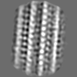
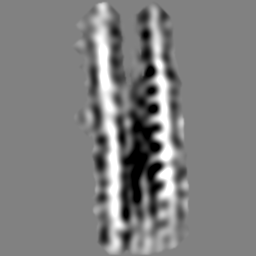
-追加マップ: Subtomogram averaging of incomplete triplet from middle centriole
| ファイル | emd_41812_additional_1.map | ||||||||||||
|---|---|---|---|---|---|---|---|---|---|---|---|---|---|
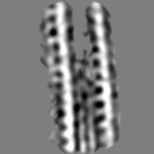
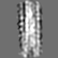
| 注釈 | Subtomogram averaging of incomplete triplet from middle centriole | ||||||||||||
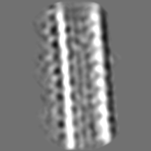
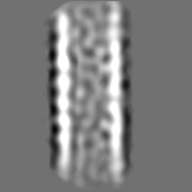
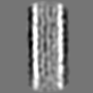
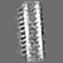
| 投影像・断面図 |
| ||||||||||||
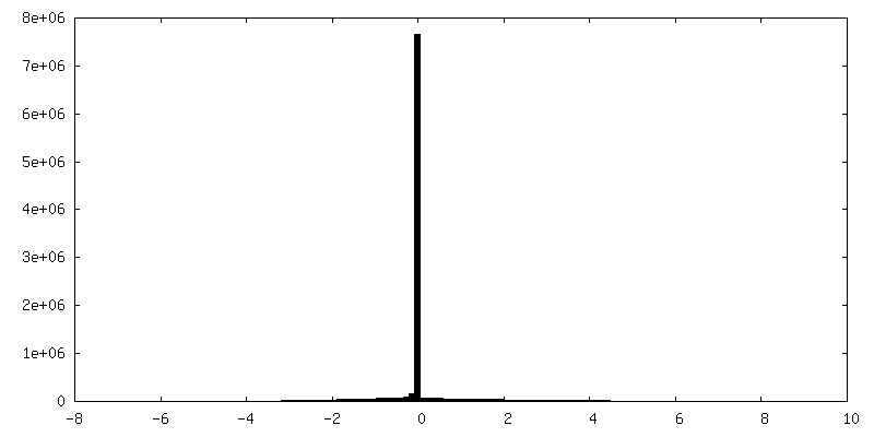
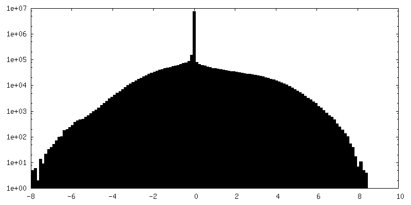
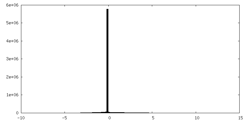
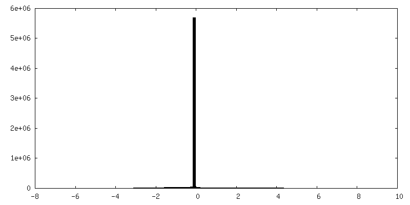
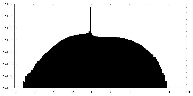
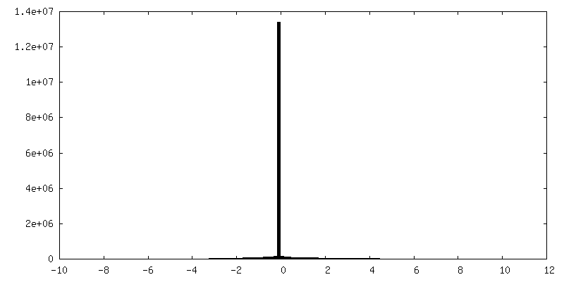
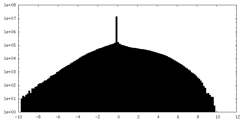
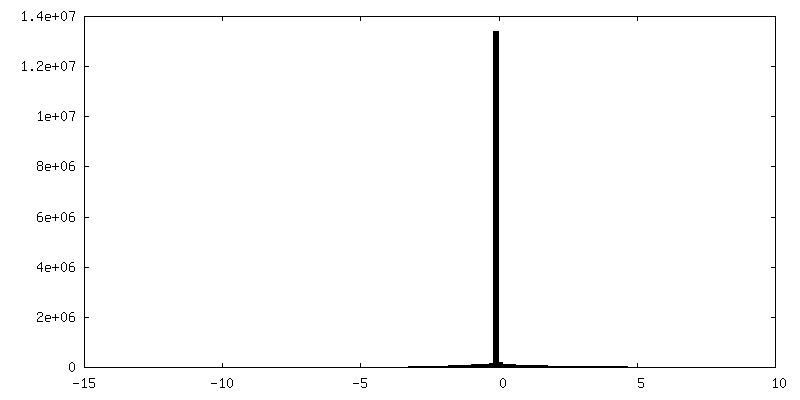
| 密度ヒストグラム |
 Z
Z Y
Y X
X

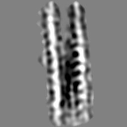
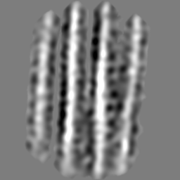
-追加マップ: Subtomogram averaging of doublets from distal centriole
| ファイル | emd_41812_additional_2.map | ||||||||||||
|---|---|---|---|---|---|---|---|---|---|---|---|---|---|
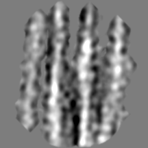
| 注釈 | Subtomogram averaging of doublets from distal centriole | ||||||||||||
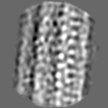
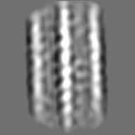
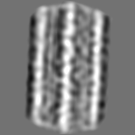
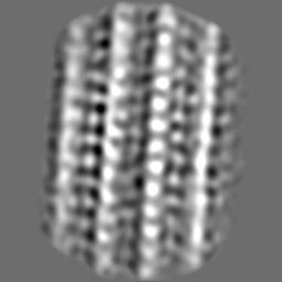
| 投影像・断面図 |
| ||||||||||||
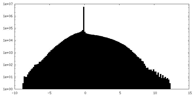
| 密度ヒストグラム |


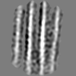
-追加マップ: Subtomogram averaging of doublets from connecting cilium
| ファイル | emd_41812_additional_3.map | ||||||||||||
|---|---|---|---|---|---|---|---|---|---|---|---|---|---|
| 注釈 | Subtomogram averaging of doublets from connecting cilium | ||||||||||||
| 投影像・断面図 |
| ||||||||||||
| 密度ヒストグラム |


-ハーフマップ: Masked odd half map
| ファイル | emd_41812_half_map_1.map | ||||||||||||
|---|---|---|---|---|---|---|---|---|---|---|---|---|---|
| 注釈 | Masked odd half map | ||||||||||||
| 投影像・断面図 |
| ||||||||||||
| 密度ヒストグラム |


-ハーフマップ: Masked even half map
| ファイル | emd_41812_half_map_2.map | ||||||||||||
|---|---|---|---|---|---|---|---|---|---|---|---|---|---|
| 注釈 | Masked even half map | ||||||||||||
| 投影像・断面図 |
| ||||||||||||
| 密度ヒストグラム |


- 試料の構成要素
-全体 : ROS(rod of outer segments)of mouse photoreceptor
| 全体 | 名称: ROS(rod of outer segments)of mouse photoreceptor |
|---|---|
| 要素 |
|
-超分子 #1: ROS(rod of outer segments)of mouse photoreceptor
| 超分子 | 名称: ROS(rod of outer segments)of mouse photoreceptor / タイプ: organelle_or_cellular_component / ID: 1 / 親要素: 0 |
|---|---|
| 由来(天然) | 生物種: |
-実験情報
-構造解析
| 手法 | クライオ電子顕微鏡法 |
|---|---|
 解析 解析 | サブトモグラム平均法 |
| 試料の集合状態 | cell |
-試料調製
| 緩衝液 | pH: 7.5 |
|---|---|
| グリッド | モデル: Quantifoil R3.5/1 / 材質: COPPER / メッシュ: 200 |
| 凍結 | 凍結剤: ETHANE |
- 電子顕微鏡法
| 顕微鏡 | FEI POLARA 300 |
|---|---|
| 撮影 | フィルム・検出器のモデル: DIRECT ELECTRON DE-12 (4k x 3k) 平均露光時間: 1.0 sec. / 平均電子線量: 10.0 e/Å2 |
| 電子線 | 加速電圧: 300 kV / 電子線源:  FIELD EMISSION GUN FIELD EMISSION GUN |
| 電子光学系 | 照射モード: FLOOD BEAM / 撮影モード: BRIGHT FIELD / 最大 デフォーカス(公称値): 1.2 µm / 最小 デフォーカス(公称値): 0.8 µm |
| 実験機器 |  モデル: Tecnai Polara / 画像提供: FEI Company |
-画像解析
| 最終 再構成 | アルゴリズム: BACK PROJECTION / 解像度のタイプ: BY AUTHOR / 解像度: 40.0 Å / 解像度の算出法: FSC 0.143 CUT-OFF / 使用したサブトモグラム数: 1800 |
|---|---|
| 抽出 | トモグラム数: 15 / 使用した粒子像数: 1800 |
| 最終 角度割当 | タイプ: PROJECTION MATCHING |
| FSC曲線 (解像度の算出) | 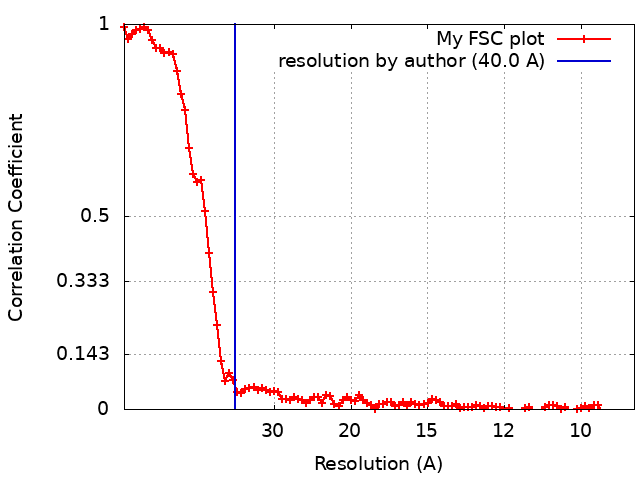 |
