 Movie
Movie Controller

[English] 日本語
 Yorodumi
- EMDB-41696: Characterization of the Chlamydomonas Flagellar Mastigoneme Filam... -
+ Open data

- Basic information
| Entry |  | |||||||||
|---|---|---|---|---|---|---|---|---|---|---|
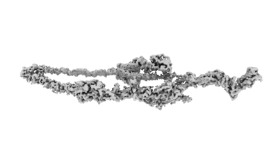
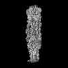
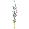
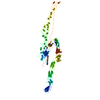
| Title | Characterization of the Chlamydomonas Flagellar Mastigoneme Filament Structure at 3.4A | |||||||||
 Map data Map data | ||||||||||
 Sample Sample |
| |||||||||
 Keywords Keywords | Mastigoneme / MST1 / flagella / Chlamydomonas / STRUCTURAL PROTEIN | |||||||||
| Biological species |   Chlamydomonas reinhardtii (plant) Chlamydomonas reinhardtii (plant) | |||||||||
| Method | single particle reconstruction / cryo EM / Resolution: 3.4 Å | |||||||||
 Authors Authors | Yue W / Kai Z | |||||||||
| Funding support |  United States, 1 items United States, 1 items
| |||||||||
 Citation Citation | Journal: J Cell Biol / Year: 2023 Title: Cryo-EM reveals how the mastigoneme assembles and responds to environmental signal changes. Authors: Yue Wang / Jun Yang / Fangheng Hu / Yuchen Yang / Kaiyao Huang / Kai Zhang /  Abstract: Mastigonemes are thread-like structures adorning the flagella of protists. In Chlamydomonas reinhardtii, filamentous mastigonemes find their roots in the flagella's distal region, associated with the ...Mastigonemes are thread-like structures adorning the flagella of protists. In Chlamydomonas reinhardtii, filamentous mastigonemes find their roots in the flagella's distal region, associated with the channel protein PKD2, implying their potential contribution to external signal sensing and flagellar motility control. Here, we present the single-particle cryo-electron microscopy structure of the mastigoneme at 3.4 Å. The filament unit, MST1, consists of nine immunoglobulin-like domains and six Sushi domains, trailed by an elastic polyproline-II helix. Our structure demonstrates that MST1 subunits are periodically assembled to form a centrosymmetric, non-polar filament. Intriguingly, numerous clustered disulfide bonds within a ladder-like spiral configuration underscore structural resilience. While defects in the mastigoneme structure did not noticeably affect general attributes of cell swimming, they did impact specific swimming properties, particularly under varied environmental conditions such as redox shifts and heightened viscosity. Our findings illuminate the potential role of mastigonemes in flagellar motility and suggest their involvement in diverse environmental responses. | |||||||||
| History |
|
- Structure visualization
| Supplemental images |
|---|
- Downloads & links
-EMDB archive
| Map data | emd_41696.map.gz | 718.8 KB |  EMDB map data format EMDB map data format | |
|---|---|---|---|---|
| Header (meta data) | emd-41696-v30.xmlemd-41696.xml | 10.9 KB 10.9 KB | Display Display | EMDB header |
| Images |  emd_41696.png emd_41696.png | 27.4 KB | ||
| Filedesc metadata | emd-41696.cif.gz | 5.2 KB | ||
| Archive directory |  http://ftp.pdbj.org/pub/emdb/structures/EMD-41696ftp://ftp.pdbj.org/pub/emdb/structures/EMD-41696 http://ftp.pdbj.org/pub/emdb/structures/EMD-41696ftp://ftp.pdbj.org/pub/emdb/structures/EMD-41696 | HTTPS FTP |
-Validation report
| Summary document | emd_41696_validation.pdf.gz | 337.6 KB | Display | EMDB validaton report |
|---|---|---|---|---|
| Full document | emd_41696_full_validation.pdf.gz | 337.2 KB | Display | |
| Data in XML | emd_41696_validation.xml.gz | 4.2 KB | Display | |
| Data in CIF | emd_41696_validation.cif.gz | 4.7 KB | Display | |
| Arichive directory | https://ftp.pdbj.org/pub/emdb/validation_reports/EMD-41696ftp://ftp.pdbj.org/pub/emdb/validation_reports/EMD-41696 | HTTPS FTP |
-Related structure data


-Links
| EMDB pages | EMDB (EBI/PDBe) / EMDataResource |
|---|
-Map
| File | Download / File: emd_41696.map.gz / Format: CCP4 / Size: 10.5 MB / Type: IMAGE STORED AS FLOATING POINT NUMBER (4 BYTES) | ||||||||||||||||||||||||||||||||||||
|---|---|---|---|---|---|---|---|---|---|---|---|---|---|---|---|---|---|---|---|---|---|---|---|---|---|---|---|---|---|---|---|---|---|---|---|---|---|
| Projections & slices | Image control
Images are generated by Spider. generated in cubic-lattice coordinate | ||||||||||||||||||||||||||||||||||||
| Voxel size | X=Y=Z: 1.33 Å | ||||||||||||||||||||||||||||||||||||
| Density |
| ||||||||||||||||||||||||||||||||||||
| Symmetry | Space group: 1 | ||||||||||||||||||||||||||||||||||||
| Details | EMDB XML:
|
 Z (Sec.)
 Y (Row.)
 X (Col.)


-Supplemental data
- Sample components
-Entire : Mastigoneme filament
| Entire | Name: Mastigoneme filament |
|---|---|
| Components |
|
-Supramolecule #1: Mastigoneme filament
| Supramolecule | Name: Mastigoneme filament / type: organelle_or_cellular_component / ID: 1 / Parent: 0 / Macromolecule list: all |
|---|---|
| Source (natural) | Organism: Chlamydomonas reinhardtii (plant) |
-Macromolecule #1: MST1
| Macromolecule | Name: MST1 / type: other / ID: 1 / Classification: other |
|---|---|
| Sequence | String: MAMPRHGRRP ARCSSNRASQ WLLLVLGVAL AASPRFLLLV EAATYTLTLD KLGPTVNPTT SDAVTFTATV ASPDSTTAVF FTLDYGDGVT AETTRTTTGA LSTTPTANLV SAGTYTVTYA SIGTKFVTLR LYDSAVAPGV LLASKTVPIY VEDSTLTATL LQSGVPRLNL ...String: MAMPRHGRRP ARCSSNRASQ WLLLVLGVAL AASPRFLLLV EAATYTLTLD KLGPTVNPTT SDAVTFTATV ASPDSTTAVF FTLDYGDGVT AETTRTTTGA LSTTPTANLV SAGTYTVTYA SIGTKFVTLR LYDSAVAPGV LLASKTVPIY VEDSTLTATL LQSGVPRLNL AFSGFKGRVS SSTANRADMW ATIQLDTAPG VFESSRIFIG IAPTASTNYD FVIPDQVYNL EGAKTTVLRI YDAPVGGTLL RTFTPAAANA VYVVDPSKYV LTLTVGPTSV TTADQVTFTQ TTTEVSYSAS ATSPILQWRF NWDDPSVVET PLAYPDALTA ASNFPTTATA VSSAAASTFR YTSTGSKNAR LRLYDGANNV IAEKIVVITV SNAGYTLALA KTTADPVTTD DTIAFSAGAK HLSSTSQVWW TIDYGAGESS PRTALTMTNV GAAAPNAIAS LSNQYTSGGT KLATLRIYDR DGVGANTGLL LASTTVTFTV TPVLYALESA VEPFSPIATV AAKWSFRIQR SKATPAGVTE SIKCAFFGAD TGTAPADLAA WLTAANGAGG LTATILPSSI PSDIISFTRT YAAAAASLQG KLQCFIGSTP LWDPYYPTPV FQVLAAAPTY TLSASVTPAV VPVDTATLWT YNIIRSVPVP AGGPSLPILC SFWDGKTGAA PTTDAGWAAL AGSANGKGTS MAPGSTTATC SFTPSYSTTG TATPTLQLIQ NSFALDAATT VGFLSPVYTA PAFATVTAAS YTISSYLNPV TPVAGGAAAV WRIVITRNAA VTASAKTLTC QMPDNGQGGS PADVTADIAV GGTTTVCVFS IAGYTTATPG PYFATVNVVD GAVTTSHITK NFTVLASGTT APTYAVTSVV SPATPVKVST PVTYTFTITR TTAVPAGGIP QPIICEFFNG EGTAPASAAA YWRVSTTIPD ADTVVAVMAP GETTTTCTFT TYYTTVSAGG FTAKLMVFGE SATAAPLLTS LSVTPSQLLA AVHSFATPMV VAAAVVAVES TTISPNYNPT TPYTNIPTYF TFTLLRDPPV PPSASSGVQF ACALYTGQNV NPASAPSAIT DAVYKTFTDV TTAVATDANY FADQQLRVVT MAPGTGRVSC TFPTLYAAAG PFSPKFFVFE YASSTVGANA LAVADTVTSL TSFTTQAAPT FITGPTNVPQ RVPLPKGFRT TCFDGYELIF SNDNYTNGVR VAVDAYPYPV GQCRKCPGGT ATMDGYRCIP CPSGYWSNEG ARECTACPAG TIAKPAALTA RAKYSIDPTT YHFVTHLAMG PESCKKCPKG YFQPNIAGTV CLPCPSGFVS TSGATGCTAC SEGTYHTDGV GTTTPGEATS LDTTDTFGSI YPIIPNTCRQ CPANTYLPLR GQAAIASMNL AAVSSATPCR PCEDGTWSKA GAAGCQKCPP GTYRNTWFSG QLGSPFITAD GVPVATTLTE LGSGCSQCPP GTYAPTFGMS VCLPCPAGTF ASAPGATACQ QCKPGTNSLM GDRTQQMALV VTNAANDFPA LRAYTISGMV AGPAYAKPIV TGPDTNFFMA GKSETCSTNL PGYYTDVDGL PIQLPCKPGT FMPFDTATAN LLDTGLTVDG TQCYTCQTGT FNDEFSQPVC KACWSGSFAS KRGLPTCEIA QPGTFTNVAA AANATFNTAT LIPTGLVKGA QAPTPCGMGY FQSSAETTTC TACAVGTYAD QAGLAACKPC QPGRYQNSIG QRVCKPCDMG TYSRYGGELC TKCPAGTVAS KTGSSQCTPC AAGFYANAPD SATSCRACPR GYYGPYSGAY ADNLGDEFEG PRGCYKCPYD FFADRPGVRQ CTACPPLDLG GGNLVEQCTE DLGSQRCKPC SLLSKPKTAR TEQSPPPPSP SPPPPPPPSP RPPSPNPPSP RPPSPAPPSP NPPPTSPPPS PPPSPPPPRP PPPPPPPPSP PPPNRSPPPP PPASSAINPG GGVNQNGDPV GHRRAILSLM EDEDAEAAQE EQAIVDVDAE MQPQDDE |
-Experimental details
-Structure determination
| Method | cryo EM |
|---|---|
 Processing Processing | single particle reconstruction |
| Aggregation state | filament |
-Sample preparation
| Buffer | pH: 7.4 |
|---|---|
| Vitrification | Cryogen name: ETHANE |
- Electron microscopy
| Microscope | FEI TITAN KRIOS |
|---|---|
| Image recording | Film or detector model: GATAN K2 QUANTUM (4k x 4k) / Average electron dose: 39.2 e/Å2 |
| Electron beam | Acceleration voltage: 300 kV / Electron source:  FIELD EMISSION GUN FIELD EMISSION GUN |
| Electron optics | Illumination mode: OTHER / Imaging mode: BRIGHT FIELD / Nominal defocus max: 2.5 µm / Nominal defocus min: 1.2 µm |
| Experimental equipment |  Model: Titan Krios / Image courtesy: FEI Company |
-Image processing
| Startup model | Type of model: NONE |
|---|---|
| Final reconstruction | Resolution.type: BY AUTHOR / Resolution: 3.4 Å / Resolution method: FSC 0.143 CUT-OFF / Number images used: 98875 |
| Initial angle assignment | Type: NOT APPLICABLE |
| Final angle assignment | Type: NOT APPLICABLE |
