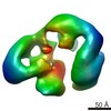
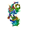
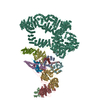
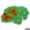
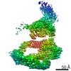
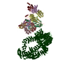
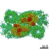
Journal: EMBO Rep / Year: 2017 Title: Architecture of the yeast Elongator complex. Authors: Maria I Dauden / Jan Kosinski / Olga Kolaj-Robin / Ambroise Desfosses / Alessandro Ori / Celine Faux / Niklas A Hoffmann / Osita F Onuma / Karin D Breunig / Martin Beck / Carsten Sachse / ...Authors: Maria I Dauden / Jan Kosinski / Olga Kolaj-Robin / Ambroise Desfosses / Alessandro Ori / Celine Faux / Niklas A Hoffmann / Osita F Onuma / Karin D Breunig / Martin Beck / Carsten Sachse / Bertrand Séraphin / Sebastian Glatt / Christoph W Müller / Abstract: The highly conserved eukaryotic Elongator complex performs specific chemical modifications on wobble base uridines of tRNAs, which are essential for proteome stability and homeostasis. The complex is ...The highly conserved eukaryotic Elongator complex performs specific chemical modifications on wobble base uridines of tRNAs, which are essential for proteome stability and homeostasis. The complex is formed by six individual subunits (Elp1-6) that are all equally important for its tRNA modification activity. However, its overall architecture and the detailed reaction mechanism remain elusive. Here, we report the structures of the fully assembled yeast Elongator and the Elp123 sub-complex solved by an integrative structure determination approach showing that two copies of the Elp1, Elp2, and Elp3 subunits form a two-lobed scaffold, which binds Elp456 asymmetrically. Our topological models are consistent with previous studies on individual subunits and further validated by complementary biochemical analyses. Our study provides a structural framework on how the tRNA modification activity is carried out by Elongator.
History
Deposition
Oct 17, 2016
-
Header (metadata) release
Oct 26, 2016
-
Map release
Dec 21, 2016
-
Update
Aug 2, 2017
-
Current status
Aug 2, 2017
Processing site: PDBe / Status: Released
-
Structure visualization
Movie
Surface view with section colored by density value
Type: NEGATIVE / Material: Uranyl Acetate Details: 3.5 ul aliquots of freshly purified Elp123 complex were applied to glow discharged carbon copper-collodion (Sigma) grids for 2 min and stained with a 1% uranyl acetate solution (w/v)
In the structure databanks used in Yorodumi, some data are registered as the other names, "COVID-19 virus" and "2019-nCoV". Here are the details of the virus and the list of structure data.
Jan 31, 2019. EMDB accession codes are about to change! (news from PDBe EMDB page)
EMDB accession codes are about to change! (news from PDBe EMDB page)
The allocation of 4 digits for EMDB accession codes will soon come to an end. Whilst these codes will remain in use, new EMDB accession codes will include an additional digit and will expand incrementally as the available range of codes is exhausted. The current 4-digit format prefixed with “EMD-” (i.e. EMD-XXXX) will advance to a 5-digit format (i.e. EMD-XXXXX), and so on. It is currently estimated that the 4-digit codes will be depleted around Spring 2019, at which point the 5-digit format will come into force.
The EM Navigator/Yorodumi systems omit the EMD- prefix.
Related info.:Q: What is EMD? / ID/Accession-code notation in Yorodumi/EM Navigator
Yorodumi is a browser for structure data from EMDB, PDB, SASBDB, etc.
This page is also the successor to EM Navigator detail page, and also detail information page/front-end page for Omokage search.
The word "yorodu" (or yorozu) is an old Japanese word meaning "ten thousand". "mi" (miru) is to see.
Related info.:EMDB / PDB / SASBDB / Comparison of 3 databanks / Yorodumi Search / Aug 31, 2016. New EM Navigator & Yorodumi / Yorodumi Papers / Jmol/JSmol / Function and homology information / Changes in new EM Navigator and Yorodumi
 Movie
Movie Controller


 Open data
 Basic information
 Map data
 Sample
 Authors
 Citation


 Structure visualization
 Movie viewer
 UCSF Chimera
 Downloads & links
 emd_4151.png
 http://ftp.pdbj.org/pub/emdb/structures/EMD-4151

 Z (Sec.)
 Y (Row.)
 X (Col.)


 Sample components
 Processing
 Electron microscopy
