National Institutes of Health/National Institute of Neurological Disorders and Stroke (NIH/NINDS)
NS112121
United States
National Institutes of Health/National Institute of Mental Health (NIH/NIMH)
MH115939
United States
National Institutes of Health/National Institute of Neurological Disorders and Stroke (NIH/NINDS)
NS105640
United States
National Institutes of Health/National Institute of Mental Health (NIH/NIMH)
R56MH122449
United States
National Institutes of Health/National Institute of General Medical Sciences (NIH/NIGMS)
R35GM142959
United States
National Institutes of Health/Office of the Director
S10OD023603
United States
Citation
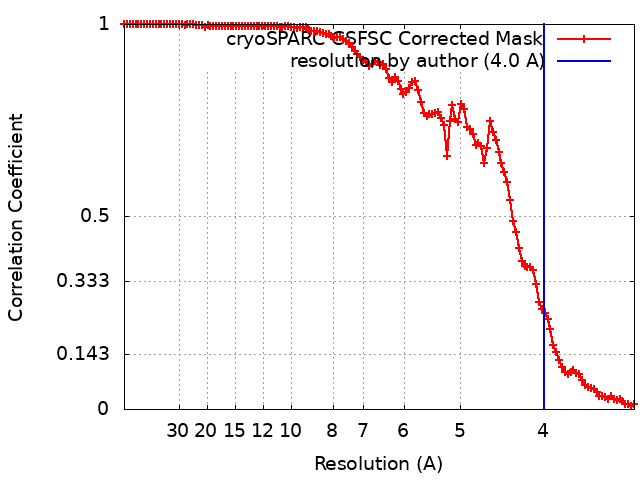
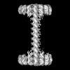
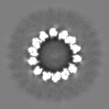
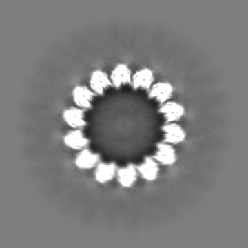
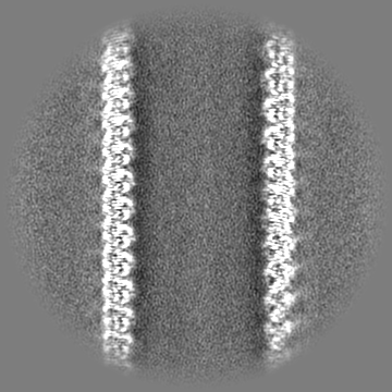
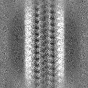
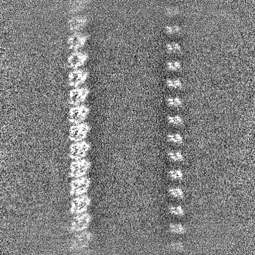
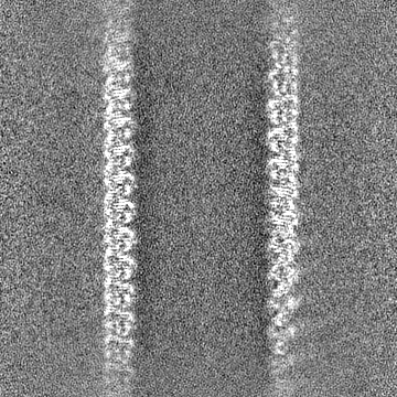
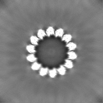
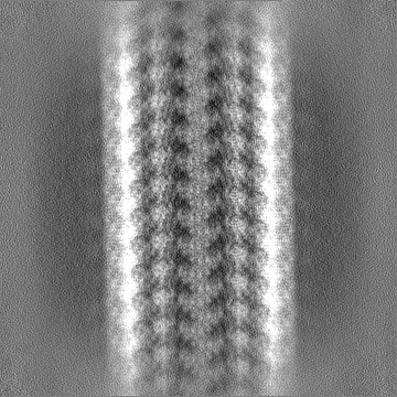
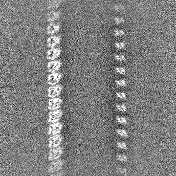
Journal: Curr Biol / Year: 2023 Title: Abl2 repairs microtubules and phase separates with tubulin to promote microtubule nucleation. Authors: Daisy Duan / Wanqing Lyu / Pengxin Chai / Shaojie Ma / Kuanlin Wu / Chunxiang Wu / Yong Xiong / Nenad Sestan / Kai Zhang / Anthony J Koleske / Abstract: Abl family kinases are evolutionarily conserved regulators of cell migration and morphogenesis. Genetic experiments in Drosophila suggest that Abl family kinases interact functionally with ...Abl family kinases are evolutionarily conserved regulators of cell migration and morphogenesis. Genetic experiments in Drosophila suggest that Abl family kinases interact functionally with microtubules to regulate axon guidance and neuronal morphogenesis. Vertebrate Abl2 binds to microtubules and promotes their plus-end elongation, both in vitro and in cells, but the molecular mechanisms by which Abl2 regulates microtubule (MT) dynamics are unclear. We report here that Abl2 regulates MT assembly via condensation and direct interactions with both the MT lattice and tubulin dimers. We find that Abl2 promotes MT nucleation, which is further facilitated by the ability of the Abl2 C-terminal half to undergo liquid-liquid phase separation (LLPS) and form co-condensates with tubulin. Abl2 binds to regions adjacent to MT damage, facilitates MT repair via fresh tubulin recruitment, and increases MT rescue frequency and lifetime. Cryo-EM analyses strongly support a model in which Abl2 engages tubulin C-terminal tails along an extended MT lattice conformation at damage sites to facilitate repair via fresh tubulin recruitment. Abl2Δ688-790, which closely mimics a naturally occurring splice isoform, retains binding to the MT lattice but does not bind tubulin, promote MT nucleation, or increase rescue frequency. In COS-7 cells, MT reassembly after nocodazole treatment is greatly slowed in Abl2 knockout COS-7 cells compared with wild-type cells, and these defects are rescued by re-expression of Abl2, but not Abl2Δ688-790. We propose that Abl2 locally concentrates tubulin to promote MT nucleation and recruits it to defects in the MT lattice to enable repair and rescue.
In the structure databanks used in Yorodumi, some data are registered as the other names, "COVID-19 virus" and "2019-nCoV". Here are the details of the virus and the list of structure data.
Jan 31, 2019. EMDB accession codes are about to change! (news from PDBe EMDB page)
EMDB accession codes are about to change! (news from PDBe EMDB page)
The allocation of 4 digits for EMDB accession codes will soon come to an end. Whilst these codes will remain in use, new EMDB accession codes will include an additional digit and will expand incrementally as the available range of codes is exhausted. The current 4-digit format prefixed with “EMD-” (i.e. EMD-XXXX) will advance to a 5-digit format (i.e. EMD-XXXXX), and so on. It is currently estimated that the 4-digit codes will be depleted around Spring 2019, at which point the 5-digit format will come into force.
The EM Navigator/Yorodumi systems omit the EMD- prefix.
Related info.:Q: What is EMD? / ID/Accession-code notation in Yorodumi/EM Navigator
Yorodumi is a browser for structure data from EMDB, PDB, SASBDB, etc.
This page is also the successor to EM Navigator detail page, and also detail information page/front-end page for Omokage search.
The word "yorodu" (or yorozu) is an old Japanese word meaning "ten thousand". "mi" (miru) is to see.
Related info.:EMDB / PDB / SASBDB / Comparison of 3 databanks / Yorodumi Search / Aug 31, 2016. New EM Navigator & Yorodumi / Yorodumi Papers / Jmol/JSmol / Function and homology information / Changes in new EM Navigator and Yorodumi
 Movie
Movie Controller


 Open data
 Basic information
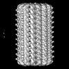
 Map data
 Sample
 Keywords
 Function and homology information
 Homo sapiens (human)
 Authors
 United States, 6 items
  Citation
 Structure visualization
 Downloads & links
 emd_41169.png
 http://ftp.pdbj.org/pub/emdb/structures/EMD-41169


 Z (Sec.)
 Y (Row.)
 X (Col.)


 Sample components
 Spodoptera frugiperda (fall armyworm)
 Processing
 Electron microscopy
 FIELD EMISSION GUN