 Movie
Movie Controller

[English] 日本語
 Yorodumi
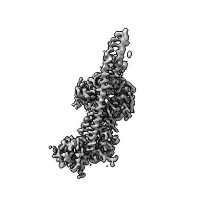
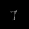
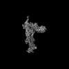
- EMDB-41056: Local refinement map (1/2) of the human PI3KC3-C1 complex (EMD-40669) -
+ Open data

- Basic information
| Entry |  | |||||||||
|---|---|---|---|---|---|---|---|---|---|---|
| Title | Local refinement map (1/2) of the human PI3KC3-C1 complex (EMD-40669) | |||||||||
 Map data Map data | Local refinement map (1/2) of the human PI3KC3-C1 complex (EMD-40669) | |||||||||
 Sample Sample |
| |||||||||
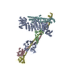
 Keywords Keywords | Autophagy / Lipid kinase / Complex / IMMUNE SYSTEM | |||||||||
| Biological species |  Homo sapiens (human) Homo sapiens (human) | |||||||||
| Method | single particle reconstruction / cryo EM / Resolution: 3.28 Å | |||||||||
 Authors Authors | Chen M / Hurley JH | |||||||||
| Funding support |  United States, 2 items United States, 2 items
| |||||||||
 Citation Citation | Journal: Nat Struct Mol Biol / Year: 2025 Title: Structure and activation of the human autophagy-initiating ULK1C:PI3KC3-C1 supercomplex. Authors: Minghao Chen / Thanh N Nguyen / Xuefeng Ren / Grace Khuu / Annan S I Cook / Yuanchang Zhao / Ahmet Yildiz / Michael Lazarou / James H Hurley /  Abstract: The Unc-51-like kinase protein kinase complex (ULK1C) is the most upstream and central player in the initiation of macroautophagy in mammals. Here, we determined the cryo-electron microscopy ...The Unc-51-like kinase protein kinase complex (ULK1C) is the most upstream and central player in the initiation of macroautophagy in mammals. Here, we determined the cryo-electron microscopy structure of the human ULK1C core at amino-acid-level resolution. We also determined a moderate-resolution structure of the ULK1C core in complex with another autophagy core complex, the class III phosphatidylinositol 3-OH kinase complex I (PI3KC3-C1). We show that the two complexes coassemble through extensive contacts between the FIP200 scaffold subunit of ULK1C and the VPS15, ATG14 and BECN1 subunits of PI3KC3-C1. The FIP200:ATG13:ULK1 core of ULK1C undergoes a rearrangement from 2:1:1 to 2:2:2 stoichiometry in the presence of PI3KC3-C1. This suggests a structural mechanism for the initiation of autophagy through formation of a ULK1C:PI3KC3-C1 supercomplex and dimerization of ULK1 on the FIP200 scaffold. #1: Journal: bioRxiv / Year: 2023Title: Structure and activation of the human autophagy-initiating ULK1C:PI3KC3-C1 supercomplex Authors: Chen M / Ren X / Cook A / Hurley JH | |||||||||
| History |
|
- Structure visualization
| Supplemental images |
|---|
- Downloads & links
-EMDB archive
| Map data | emd_41056.map.gz | 164.7 MB |  EMDB map data format EMDB map data format | |
|---|---|---|---|---|
| Header (meta data) | emd-41056-v30.xmlemd-41056.xml | 18.4 KB 18.4 KB | Display Display | EMDB header |
| Images |  emd_41056.png emd_41056.png | 54.9 KB | ||
| Filedesc metadata | emd-41056.cif.gz | 4.6 KB | ||
| Others | emd_41056_half_map_1.map.gzemd_41056_half_map_2.map.gz | 164.9 MB 164.9 MB | ||
| Archive directory |  http://ftp.pdbj.org/pub/emdb/structures/EMD-41056ftp://ftp.pdbj.org/pub/emdb/structures/EMD-41056 http://ftp.pdbj.org/pub/emdb/structures/EMD-41056ftp://ftp.pdbj.org/pub/emdb/structures/EMD-41056 | HTTPS FTP |
-Related structure data


-Links
| EMDB pages | EMDB (EBI/PDBe) / EMDataResource |
|---|
-Map
| File | Download / File: emd_41056.map.gz / Format: CCP4 / Size: 178 MB / Type: IMAGE STORED AS FLOATING POINT NUMBER (4 BYTES) | ||||||||||||||||||||||||||||||||||||
|---|---|---|---|---|---|---|---|---|---|---|---|---|---|---|---|---|---|---|---|---|---|---|---|---|---|---|---|---|---|---|---|---|---|---|---|---|---|
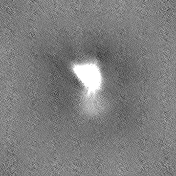
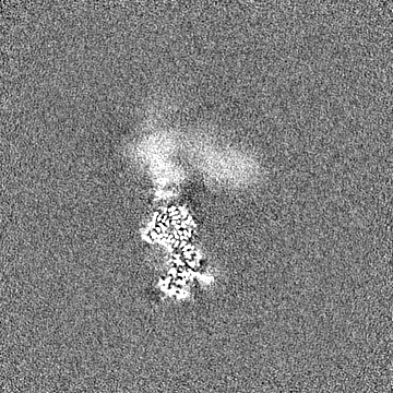
| Annotation | Local refinement map (1/2) of the human PI3KC3-C1 complex (EMD-40669) | ||||||||||||||||||||||||||||||||||||
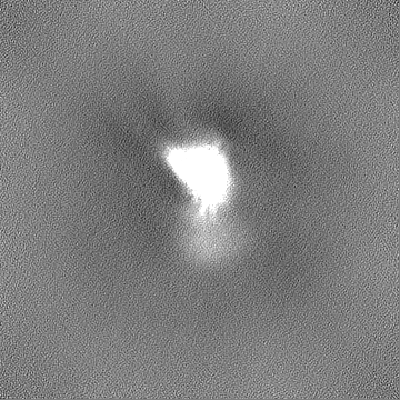
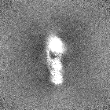
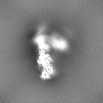
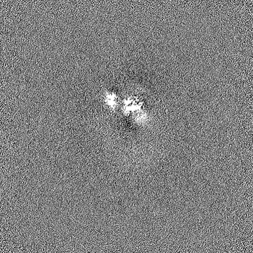
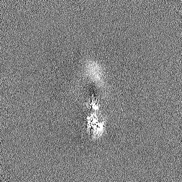
| Projections & slices | Image control
Images are generated by Spider. | ||||||||||||||||||||||||||||||||||||
| Voxel size | X=Y=Z: 1.115 Å | ||||||||||||||||||||||||||||||||||||
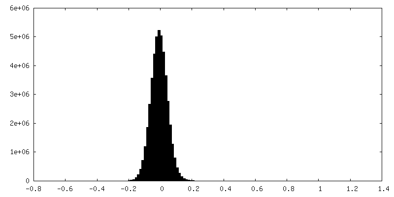
| Density |
| ||||||||||||||||||||||||||||||||||||
| Symmetry | Space group: 1 | ||||||||||||||||||||||||||||||||||||
| Details | EMDB XML:
|
 Z (Sec.)
 Y (Row.)
 X (Col.)


-Supplemental data
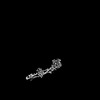
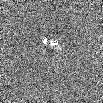
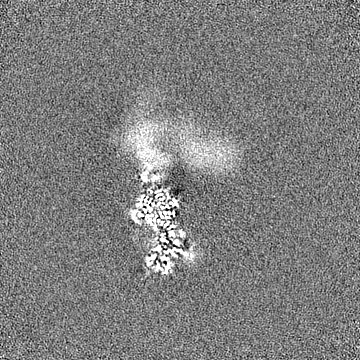
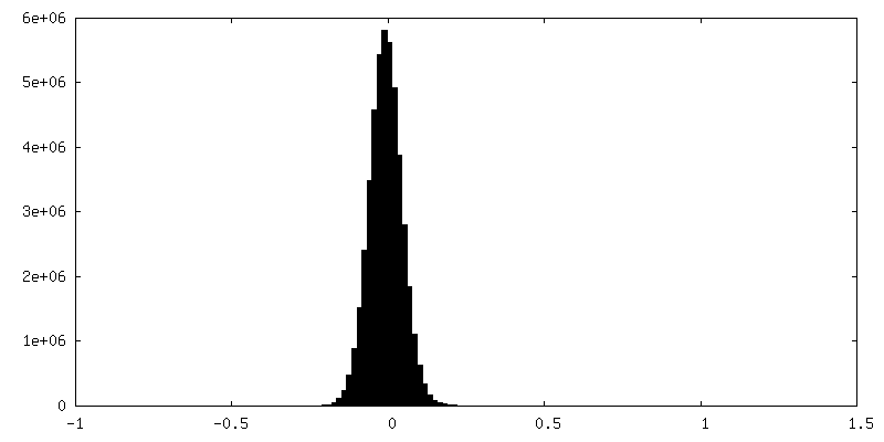
-Half map: The according half map (1/2)
| File | emd_41056_half_map_1.map | ||||||||||||
|---|---|---|---|---|---|---|---|---|---|---|---|---|---|
| Annotation | The according half map (1/2) | ||||||||||||
| Projections & Slices |
| ||||||||||||
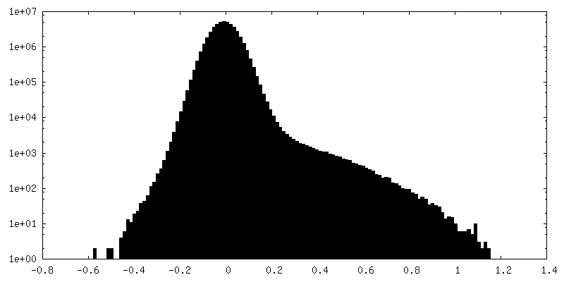
| Density Histograms |


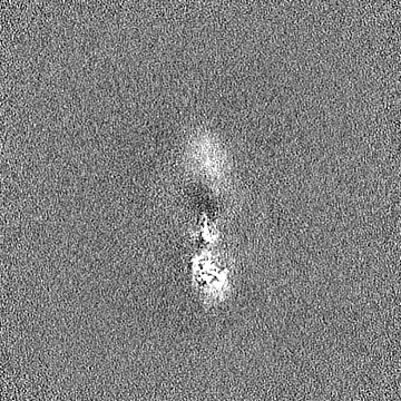
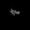
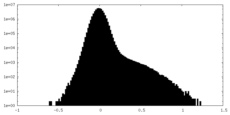
-Half map: The according half map (2/2)
| File | emd_41056_half_map_2.map | ||||||||||||
|---|---|---|---|---|---|---|---|---|---|---|---|---|---|
| Annotation | The according half map (2/2) | ||||||||||||
| Projections & Slices |
| ||||||||||||
| Density Histograms |
- Sample components
-Entire : Human autophagy initiation PI3KC3-C1 complex
| Entire | Name: Human autophagy initiation PI3KC3-C1 complex |
|---|---|
| Components |
|
-Supramolecule #1: Human autophagy initiation PI3KC3-C1 complex
| Supramolecule | Name: Human autophagy initiation PI3KC3-C1 complex / type: complex / ID: 1 / Parent: 0 / Macromolecule list: #1-#4 |
|---|---|
| Source (natural) | Organism: Homo sapiens (human) |
| Molecular weight | Theoretical: 362 KDa |
-Experimental details
-Structure determination
| Method | cryo EM |
|---|---|
 Processing Processing | single particle reconstruction |
| Aggregation state | particle |
-Sample preparation
| Concentration | 0.25 mg/mL | |||||||||||||||
|---|---|---|---|---|---|---|---|---|---|---|---|---|---|---|---|---|
| Buffer | pH: 7.4 Component:
| |||||||||||||||
| Grid | Model: Quantifoil R1.2/1.3 / Material: COPPER / Mesh: 300 / Support film - Material: CARBON / Support film - topology: HOLEY / Details: 25 mA | |||||||||||||||
| Vitrification | Cryogen name: ETHANE / Chamber humidity: 100 % / Chamber temperature: 277 K / Instrument: FEI VITROBOT MARK IV |
- Electron microscopy
| Microscope | FEI TALOS ARCTICA |
|---|---|
| Image recording | Film or detector model: GATAN K3 (6k x 4k) / Number grids imaged: 1 / Number real images: 2243 / Average electron dose: 50.0 e/Å2 |
| Electron beam | Acceleration voltage: 200 kV / Electron source:  FIELD EMISSION GUN FIELD EMISSION GUN |
| Electron optics | C2 aperture diameter: 50.0 µm / Illumination mode: FLOOD BEAM / Imaging mode: BRIGHT FIELD / Cs: 2.7 mm / Nominal defocus max: 2.0 µm / Nominal defocus min: 0.8 µm / Nominal magnification: 36000 |
| Sample stage | Cooling holder cryogen: NITROGEN |
| Experimental equipment |  Model: Talos Arctica / Image courtesy: FEI Company |
+Image processing
-Atomic model buiding 1
| Initial model | Chain - Source name: AlphaFold / Chain - Initial model type: in silico model |
|---|---|
| Refinement | Space: REAL / Protocol: AB INITIO MODEL |
