[English] 日本語
 Yorodumi
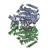
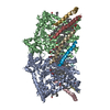
- EMDB-41044: Structure of mechanically activated ion channel OSCA2.3 in peptidiscs -
+ Open data
- Basic information
| Entry |  | |||||||||
|---|---|---|---|---|---|---|---|---|---|---|
| Title | Structure of mechanically activated ion channel OSCA2.3 in peptidiscs | |||||||||
 Map data Map data | OSCA2.3 in peptidisc postprocessed with LocalDeblur | |||||||||
 Sample Sample |
| |||||||||
 Keywords Keywords | mechanically activated ion channel / MEMBRANE PROTEIN | |||||||||
| Function / homology |  Function and homology information Function and homology informationmechanosensitive monoatomic ion channel activity / calcium-activated cation channel activity / membrane Similarity search - Function | |||||||||
| Biological species |  | |||||||||
| Method | single particle reconstruction / cryo EM / Resolution: 2.7 Å | |||||||||
 Authors Authors | Jojoa-Cruz S / Burendei B / Lee WH / Ward AB | |||||||||
| Funding support |  United States, 1 items United States, 1 items
| |||||||||
 Citation Citation |  Journal: Structure / Year: 2024 Journal: Structure / Year: 2024Title: Structure of mechanically activated ion channel OSCA2.3 reveals mobile elements in the transmembrane domain. Authors: Sebastian Jojoa-Cruz / Batuujin Burendei / Wen-Hsin Lee / Andrew B Ward /  Abstract: Members of the OSCA/TMEM63 family are mechanically activated ion channels and structures of some OSCA members have revealed the architecture of these channels and structural features that are ...Members of the OSCA/TMEM63 family are mechanically activated ion channels and structures of some OSCA members have revealed the architecture of these channels and structural features that are potentially involved in mechanosensation. However, these structures are all in a similar state and information about the motion of different elements of the structure is limited, preventing a deeper understanding of how these channels work. Here, we used cryoelectron microscopy to determine high-resolution structures of Arabidopsis thaliana OSCA1.2 and OSCA2.3 in peptidiscs. The structure of OSCA1.2 matches previous structures of the same protein in different environments. Yet, in OSCA2.3, the TM6a-TM7 linker adopts a different conformation that constricts the pore on its cytoplasmic side. Furthermore, coevolutionary sequence analysis uncovered a conserved interaction between the TM6a-TM7 linker and the beam-like domain (BLD). Our results reveal conformational heterogeneity and differences in conserved interactions between the TMD and BLD among members of the OSCA family. | |||||||||
| History |
|
- Structure visualization
| Supplemental images |
|---|
- Downloads & links
-EMDB archive
| Map data |  emd_41044.map.gz emd_41044.map.gz | 22.4 MB |  EMDB map data format EMDB map data format | |
|---|---|---|---|---|
| Header (meta data) |  emd-41044-v30.xml emd-41044-v30.xml emd-41044.xml emd-41044.xml | 20.8 KB 20.8 KB | Display Display |  EMDB header EMDB header |
| FSC (resolution estimation) |  emd_41044_fsc.xml emd_41044_fsc.xml | 7.1 KB | Display |  FSC data file FSC data file |
| Images |  emd_41044.png emd_41044.png | 81.7 KB | ||
| Filedesc metadata |  emd-41044.cif.gz emd-41044.cif.gz | 6.5 KB | ||
| Others |  emd_41044_additional_1.map.gz emd_41044_additional_1.map.gz emd_41044_half_map_1.map.gz emd_41044_half_map_1.map.gz emd_41044_half_map_2.map.gz emd_41044_half_map_2.map.gz | 26.6 MB 22.5 MB 22.5 MB | ||
| Archive directory |  http://ftp.pdbj.org/pub/emdb/structures/EMD-41044 http://ftp.pdbj.org/pub/emdb/structures/EMD-41044 ftp://ftp.pdbj.org/pub/emdb/structures/EMD-41044 ftp://ftp.pdbj.org/pub/emdb/structures/EMD-41044 | HTTPS FTP |
-Validation report
| Summary document |  emd_41044_validation.pdf.gz emd_41044_validation.pdf.gz | 785.1 KB | Display |  EMDB validaton report EMDB validaton report |
|---|---|---|---|---|
| Full document |  emd_41044_full_validation.pdf.gz emd_41044_full_validation.pdf.gz | 784.7 KB | Display | |
| Data in XML |  emd_41044_validation.xml.gz emd_41044_validation.xml.gz | 12.5 KB | Display | |
| Data in CIF |  emd_41044_validation.cif.gz emd_41044_validation.cif.gz | 17.4 KB | Display | |
| Arichive directory |  https://ftp.pdbj.org/pub/emdb/validation_reports/EMD-41044 https://ftp.pdbj.org/pub/emdb/validation_reports/EMD-41044 ftp://ftp.pdbj.org/pub/emdb/validation_reports/EMD-41044 ftp://ftp.pdbj.org/pub/emdb/validation_reports/EMD-41044 | HTTPS FTP |
-Related structure data
| Related structure data |  8t57MC  8t56C M: atomic model generated by this map C: citing same article ( |
|---|---|
| Similar structure data | Similarity search - Function & homology  F&H Search F&H Search |
- Links
Links
| EMDB pages |  EMDB (EBI/PDBe) / EMDB (EBI/PDBe) /  EMDataResource EMDataResource |
|---|
- Map
Map
| File |  Download / File: emd_41044.map.gz / Format: CCP4 / Size: 30.5 MB / Type: IMAGE STORED AS FLOATING POINT NUMBER (4 BYTES) Download / File: emd_41044.map.gz / Format: CCP4 / Size: 30.5 MB / Type: IMAGE STORED AS FLOATING POINT NUMBER (4 BYTES) | ||||||||||||||||||||||||||||||||||||
|---|---|---|---|---|---|---|---|---|---|---|---|---|---|---|---|---|---|---|---|---|---|---|---|---|---|---|---|---|---|---|---|---|---|---|---|---|---|
| Annotation | OSCA2.3 in peptidisc postprocessed with LocalDeblur | ||||||||||||||||||||||||||||||||||||
| Projections & slices | Image control
Images are generated by Spider. | ||||||||||||||||||||||||||||||||||||
| Voxel size | X=Y=Z: 1.03 Å | ||||||||||||||||||||||||||||||||||||
| Density |
| ||||||||||||||||||||||||||||||||||||
| Symmetry | Space group: 1 | ||||||||||||||||||||||||||||||||||||
| Details | EMDB XML:
|
-Supplemental data
-Additional map: OSCA2.3 in peptidisc postprocessed with DeepEMhancer "highRes" model
| File | emd_41044_additional_1.map | ||||||||||||
|---|---|---|---|---|---|---|---|---|---|---|---|---|---|
| Annotation | OSCA2.3 in peptidisc postprocessed with DeepEMhancer "highRes" model | ||||||||||||
| Projections & Slices |
| ||||||||||||
| Density Histograms |
-Half map: OSCA2.3 in peptidisc half map 1
| File | emd_41044_half_map_1.map | ||||||||||||
|---|---|---|---|---|---|---|---|---|---|---|---|---|---|
| Annotation | OSCA2.3 in peptidisc half map 1 | ||||||||||||
| Projections & Slices |
| ||||||||||||
| Density Histograms |
-Half map: OSCA2.3 in peptidisc half map 2
| File | emd_41044_half_map_2.map | ||||||||||||
|---|---|---|---|---|---|---|---|---|---|---|---|---|---|
| Annotation | OSCA2.3 in peptidisc half map 2 | ||||||||||||
| Projections & Slices |
| ||||||||||||
| Density Histograms |
- Sample components
-Entire : OSCA2.3 in peptidisc
| Entire | Name: OSCA2.3 in peptidisc |
|---|---|
| Components |
|
-Supramolecule #1: OSCA2.3 in peptidisc
| Supramolecule | Name: OSCA2.3 in peptidisc / type: complex / ID: 1 / Parent: 0 / Macromolecule list: #1 |
|---|---|
| Source (natural) | Organism:  |
| Molecular weight | Theoretical: 159 KDa |
-Macromolecule #1: CSC1-like protein HYP1
| Macromolecule | Name: CSC1-like protein HYP1 / type: protein_or_peptide / ID: 1 Details: The last 10 residues (GTGTLEVLFQ) are leftover of a linker and protease site. Number of copies: 2 / Enantiomer: LEVO |
|---|---|
| Source (natural) | Organism:  |
| Molecular weight | Theoretical: 80.848078 KDa |
| Recombinant expression | Organism:  Homo sapiens (human) Homo sapiens (human) |
| Sequence | String: MLLSALLTSV GINLGLCFLF FTLYSILRKQ PSNVTVYGPR LVKKDGKSQQ SNEFNLERLL PTAGWVKRAL EPTNDEILSN LGLDALVFI RVFVFSIRVF SFASVVGIFI LLPVNYMGTE FEEFFDLPKK SMDNFSISNV NDGSNKLWIH FCAIYIFTAV V CSLLYYEH ...String: MLLSALLTSV GINLGLCFLF FTLYSILRKQ PSNVTVYGPR LVKKDGKSQQ SNEFNLERLL PTAGWVKRAL EPTNDEILSN LGLDALVFI RVFVFSIRVF SFASVVGIFI LLPVNYMGTE FEEFFDLPKK SMDNFSISNV NDGSNKLWIH FCAIYIFTAV V CSLLYYEH KYILTKRIAH LYSSKPQPQE FTVLVSGVPL VSGNSISETV ENFFREYHSS SYLSHIVVHR TDKLKVLMND AE KLYKKLT RVKSGSISRQ KSRWGGFLGM FGNNVDVVDH YQKKLDKLED DMRLKQSLLA GEEVPAAFVS FRTRHGAAIA TNI QQGIDP TQWLTEAAPE PEDVHWPFFT ASFVRRWISN VVVLVAFVAL LILYIVPVVL VQGLANLHQL ETWFPFLKGI LNMK IVSQV ITGYLPSLIF QLFLLIVPPI MLLLSSMQGF ISHSQIEKSA CIKLLIFTVW NSFFANVLSG SALYRVNVFL EPKTI PRVL AAAVPAQASF FVSYVVTSGW TGLSSEILRL VPLLWSFITK LFGKEDDKEF EVPSTPFCQE IPRILFFGLL GITYFF LSP LILPFLLVYY CLGYIIYRNQ LLNVYAAKYE TGGKFWPIVH SYTIFSLVLM HIIAVGLFGL KELPVASSLT IPLPVLT VL FSIYCQRRFL PNFKSYPTQC LVNKDKADER EQNMSEFYSE LVVAYRDPAL SASQDSRDIS PGTGTLEVLF Q UniProtKB: Hyperosmolality-gated Ca2+ permeable channel 2.3 |
-Macromolecule #2: CHOLESTEROL
| Macromolecule | Name: CHOLESTEROL / type: ligand / ID: 2 / Number of copies: 4 / Formula: CLR |
|---|---|
| Molecular weight | Theoretical: 386.654 Da |
| Chemical component information |  ChemComp-CLR: |
-Macromolecule #3: PALMITIC ACID
| Macromolecule | Name: PALMITIC ACID / type: ligand / ID: 3 / Number of copies: 16 / Formula: PLM |
|---|---|
| Molecular weight | Theoretical: 256.424 Da |
| Chemical component information |  ChemComp-PLM: |
-Experimental details
-Structure determination
| Method | cryo EM |
|---|---|
 Processing Processing | single particle reconstruction |
| Aggregation state | particle |
- Sample preparation
| Concentration | 3.6 mg/mL | ||||||||||||
|---|---|---|---|---|---|---|---|---|---|---|---|---|---|
| Buffer | pH: 8 Component:
| ||||||||||||
| Grid | Material: GOLD / Mesh: 300 | ||||||||||||
| Vitrification | Cryogen name: ETHANE / Chamber humidity: 100 % / Chamber temperature: 277 K / Instrument: FEI VITROBOT MARK IV |
- Electron microscopy
| Microscope | FEI TITAN KRIOS |
|---|---|
| Image recording | Film or detector model: GATAN K2 SUMMIT (4k x 4k) / Detector mode: COUNTING / Digitization - Dimensions - Width: 3710 pixel / Digitization - Dimensions - Height: 3838 pixel / Number grids imaged: 3 / Number real images: 5323 / Average electron dose: 50.0 e/Å2 |
| Electron beam | Acceleration voltage: 300 kV / Electron source:  FIELD EMISSION GUN FIELD EMISSION GUN |
| Electron optics | C2 aperture diameter: 70.0 µm / Illumination mode: FLOOD BEAM / Imaging mode: BRIGHT FIELD / Cs: 2.7 mm / Nominal defocus max: 1.5 µm / Nominal defocus min: 0.6 µm / Nominal magnification: 29000 |
| Sample stage | Specimen holder model: FEI TITAN KRIOS AUTOGRID HOLDER / Cooling holder cryogen: NITROGEN |
| Experimental equipment |  Model: Titan Krios / Image courtesy: FEI Company |
 Movie
Movie Controller


 Z (Sec.)
 Y (Row.)
 X (Col.)


