 Movie
Movie Controller


+ Open data

- Basic information
| Entry | Database: EMDB / ID: EMD-4066 | |||||||||
|---|---|---|---|---|---|---|---|---|---|---|
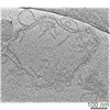
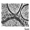
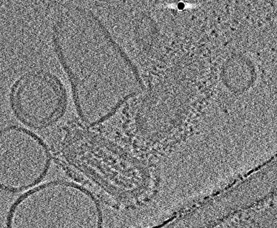
| Title | Influenza virus membrane fusion | |||||||||
 Map data Map data | ||||||||||
 Sample Sample |
| |||||||||
| Biological species |  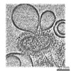 unidentified influenza virus unidentified influenza virus | |||||||||
| Method | electron tomography / cryo EM | |||||||||
 Authors Authors | Rosenthal PB / Calder LJ | |||||||||
 Citation Citation | Journal: Nat Struct Mol Biol / Year: 2016 Title: Cryomicroscopy provides structural snapshots of influenza virus membrane fusion. Authors: Lesley J Calder / Peter B Rosenthal /  Abstract: The lipid-enveloped influenza virus enters host cells during infection by binding cell-surface receptors and, after receptor-mediated endocytosis, fusing with the membrane of the endosome and ...The lipid-enveloped influenza virus enters host cells during infection by binding cell-surface receptors and, after receptor-mediated endocytosis, fusing with the membrane of the endosome and delivering the viral genome and transcription machinery into the host cell. These events are mediated by the hemagglutinin (HA) surface glycoprotein. At the low pH of the endosome, an irreversible conformational change in the HA, including the exposure of the hydrophobic fusion peptide, activates membrane fusion. Here we used electron cryomicroscopy and cryotomography to image the fusion of influenza virus with target membranes at low pH. We visualized structural intermediates of HA and their interactions with membranes during the course of membrane fusion as well as ultrastructural changes in the virus that accompany membrane fusion. Our observations are relevant to a wide range of protein-mediated membrane-fusion processes and demonstrate how dynamic membrane events may be studied by cryomicroscopy. | |||||||||
| History |
|
- Structure visualization
| Movie |
 Movie viewer Movie viewer |
|---|---|
| Structure viewer | EM map: SurfViewMolmilJmol/JSmol |
| Supplemental images |

- Downloads & links
-EMDB archive
| Map data | emd_4066.map.gz | 161.6 MB | EMDB map data format | |
|---|---|---|---|---|
| Header (meta data) | emd-4066-v30.xmlemd-4066.xml | 7.8 KB 7.8 KB | Display Display | EMDB header |
| Images | 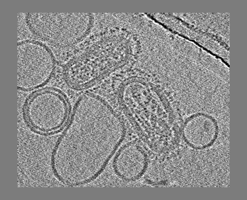 emd_4066.png emd_4066.png | 137.2 KB | ||
| Archive directory |  http://ftp.pdbj.org/pub/emdb/structures/EMD-4066ftp://ftp.pdbj.org/pub/emdb/structures/EMD-4066 http://ftp.pdbj.org/pub/emdb/structures/EMD-4066ftp://ftp.pdbj.org/pub/emdb/structures/EMD-4066 | HTTPS FTP |
-Related structure data
-Links
| EMDB pages | EMDB (EBI/PDBe) / EMDataResource |
|---|
-Map
| File | Download / File: emd_4066.map.gz / Format: CCP4 / Size: 521.6 MB / Type: IMAGE STORED AS FLOATING POINT NUMBER (4 BYTES) | ||||||||||||||||||||||||||||||||||||||||||||||||||||||||||||
|---|---|---|---|---|---|---|---|---|---|---|---|---|---|---|---|---|---|---|---|---|---|---|---|---|---|---|---|---|---|---|---|---|---|---|---|---|---|---|---|---|---|---|---|---|---|---|---|---|---|---|---|---|---|---|---|---|---|---|---|---|---|
| Projections & slices | Image control
Images are generated by Spider. generated in cubic-lattice coordinate | ||||||||||||||||||||||||||||||||||||||||||||||||||||||||||||
| Voxel size | X=Y=Z: 4.3 Å | ||||||||||||||||||||||||||||||||||||||||||||||||||||||||||||
| Density |
| ||||||||||||||||||||||||||||||||||||||||||||||||||||||||||||
| Symmetry | Space group: 1 | ||||||||||||||||||||||||||||||||||||||||||||||||||||||||||||
| Details | EMDB XML:
CCP4 map header:
| ||||||||||||||||||||||||||||||||||||||||||||||||||||||||||||
 Z (Sec.)
 Y (Row.)
 X (Col.)


-Supplemental data
- Sample components
-Entire : unidentified influenza virus
| Entire | Name: unidentified influenza virus |
|---|---|
| Components |
|
-Supramolecule #1: unidentified influenza virus
| Supramolecule | Name: unidentified influenza virus / type: virus / ID: 1 / Parent: 0 / Macromolecule list: #1 / NCBI-ID: 11309 / Sci species name: unidentified influenza virus / Sci species strain: A/Udorn/72 / Virus type: VIRION / Virus isolate: STRAIN / Virus enveloped: Yes / Virus empty: No |
|---|
-Experimental details
-Structure determination
| Method | cryo EM |
|---|---|
 Processing Processing | electron tomography |
| Aggregation state | particle |
-Sample preparation
| Buffer | pH: 5 Details: Sample was incubated at pH5 for 1min and then neutralised |
|---|---|
| Vitrification | Cryogen name: ETHANE |
| Sectioning | Other: NO SECTIONING |
| Fiducial marker | Manufacturer: British Biocell / Diameter: 10 nm |
- Electron microscopy
| Microscope | FEI POLARA 300 |
|---|---|
| Image recording | Film or detector model: TVIPS TEMCAM-F224 (2k x 2k) / Average electron dose: 2.0 e/Å2 |
| Electron beam | Acceleration voltage: 200 kV / Electron source:  FIELD EMISSION GUN FIELD EMISSION GUN |
| Electron optics | Illumination mode: FLOOD BEAM / Imaging mode: BRIGHT FIELD |
| Experimental equipment |  Model: Tecnai Polara / Image courtesy: FEI Company |
-Image processing
| Final reconstruction | Algorithm: SIMULTANEOUS ITERATIVE (SIRT) / Software - Name: IMOD / Number images used: 40 |
|---|
