National Institutes of Health/National Institute of General Medical Sciences (NIH/NIGMS)
P41GM136508
米国
Department of Defense (DOD, United States)
HDTRA1-21-1-0004
米国
Howard Hughes Medical Institute (HHMI)
米国
引用
ジャーナル: IUCrJ / 年: 2023 タイトル: Design and implementation of suspended drop crystallization. 著者: Cody Gillman / William J Nicolas / Michael W Martynowycz / Tamir Gonen / 要旨: In this work, a novel crystal growth method termed suspended drop crystallization has been developed. Unlike traditional methods, this technique involves mixing protein and precipitant directly on an ...In this work, a novel crystal growth method termed suspended drop crystallization has been developed. Unlike traditional methods, this technique involves mixing protein and precipitant directly on an electron microscopy grid without any additional support layers. The grid is then suspended within a crystallization chamber designed in-house, allowing for vapor diffusion to occur from both sides of the drop. A UV-transparent window above and below the grid enables the monitoring of crystal growth via light, UV or fluorescence microscopy. Once crystals have formed, the grid can be removed and utilized for X-ray crystallography or microcrystal electron diffraction (MicroED) directly without having to manipulate the crystals. To demonstrate the efficacy of this method, crystals of the enzyme proteinase K were grown and its structure was determined by MicroED following focused ion beam/scanning electron microscopy milling to render the sample thin enough for cryoEM. Suspended drop crystallization overcomes many of the challenges associated with sample preparation, providing an alternative workflow for crystals embedded in viscous media, sensitive to mechanical stress and/or subject to preferred orientation on electron microscopy grids.
 ムービー
ムービー コントローラー


 データを開く
 基本情報
基本情報
 マップデータ
 試料
試料 キーワード
キーワード 機能・相同性情報
 Parengyodontium album (菌類)
 データ登録者
 米国, 3件
  引用
引用 構造の表示
構造の表示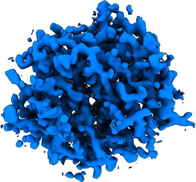
 ダウンロードとリンク
 emd_40351.png
 http://ftp.pdbj.org/pub/emdb/structures/EMD-40351
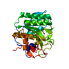


 X (Sec.)
 Y (Row.)
 Z (Col.)


 試料の構成要素

 解析
解析 電子顕微鏡法
 FIELD EMISSION GUN
